Single-cell analysis is a method that offers a brand new opportunity to study the body’s organisms and tissue, right down to gene activity in an individual cell. Sten Linnarsson and his team want to understand why certain cells specialize in certain functions in specific organs. And at the center of his research lies the most complex organ of all – the brain.
Project Grant 2015
Cis-regulatory logic of the mouse brain transcriptome
Principal investigator:
Sten Linnarsson, Professor of Molecular System Biology, specializing in transcriptomics
Institution:
Karolinska Institutet
Grant in SEK:
SEK 26.8 million
“I want to build a map of how the brain develops normally, so it will then be possible to study the course of various diseases. The impetus for this research is not the applications, but learning how the brain develops and works.”
His main interest is in embryonic development – how the brain develops at the fetal stage. Some conditions originating in the brain as early as the fetal stage are developmental impairment, schizophrenia and autism.
“If we are to understand the brain, we must first find and sort all its components, so we can then examine and map how they work together.”
One of the pioneers
Single-cell analysis is a fairly new method, and Professor Linnarsson is one of its pioneers. A major breakthrough for the method came around 2013, when it was found to be reliable, and it is now used in every area of biological research.
“Earlier it was not possible to study genes in individual cells. A gene was either studied through a section of tissue under a microscope, or by taking a tissue sample, blending it, and obtaining an average value for various constituents.”
He draws a parallel in which earlier samples were like a fruit salad pressed to make a juice. With single-cell analysis the separate parts can be studied individually, sorted into piles according to type, and examined to see how they fit together.
The method makes it possible to apply various strategies. A disease such as MS, for example, which affects myelin in the brain, can be studied, or the focus may instead be on understanding brain structure.
“There are two parts to our project – we want to map cell types in the brain, and the areas where they are found, and we also want to understand the mechanisms causing cells to specialize in various functions. Why are some genes active, producing certain enzymes in certain cells? There is a regulatory mechanism during embryonic development to ensure that we get the right cells in the right place.”
Map of the cerebral cortex
Professor Linnarsson and his team have already published a study in Science, in which they have made a map of the brain using single-cell analysis, showing which genes are active in the various cells. They studied just over 3,000 cells, one by one, from the cerebral cortex of mice, and were also able to identify previously unknown cell types. With the help of funding from the Knut and Alice Wallenberg Foundation, they will now go on to map the remainder of the cerebral cortex and other parts of the brain.
“We hope to be able to make an atlas of the whole brain. The thing is that even though we can now study individual cells, we don’t know where they come from. We don’t know about the architecture. When we know which cells there are, and what their function is, we can go back to the tissue, color in the genes, and see where they are located, thereby also gaining an insight into the importance of the surroundings.
To achieve this they will be using a mouse model system.
“Comparative studies have shown great similarities between mice and humans,” Linnarsson comments.
The team is also studying a kind of regulatory element, known as “enhancers”, which are DNA sequences controlling gene activity. They may be likened to switches that can turn genes on and off so they prevent each other from doing what they are supposed to do. This may trigger disease, including tumor growth. Many types of cancer are caused by mutations in enhancers.
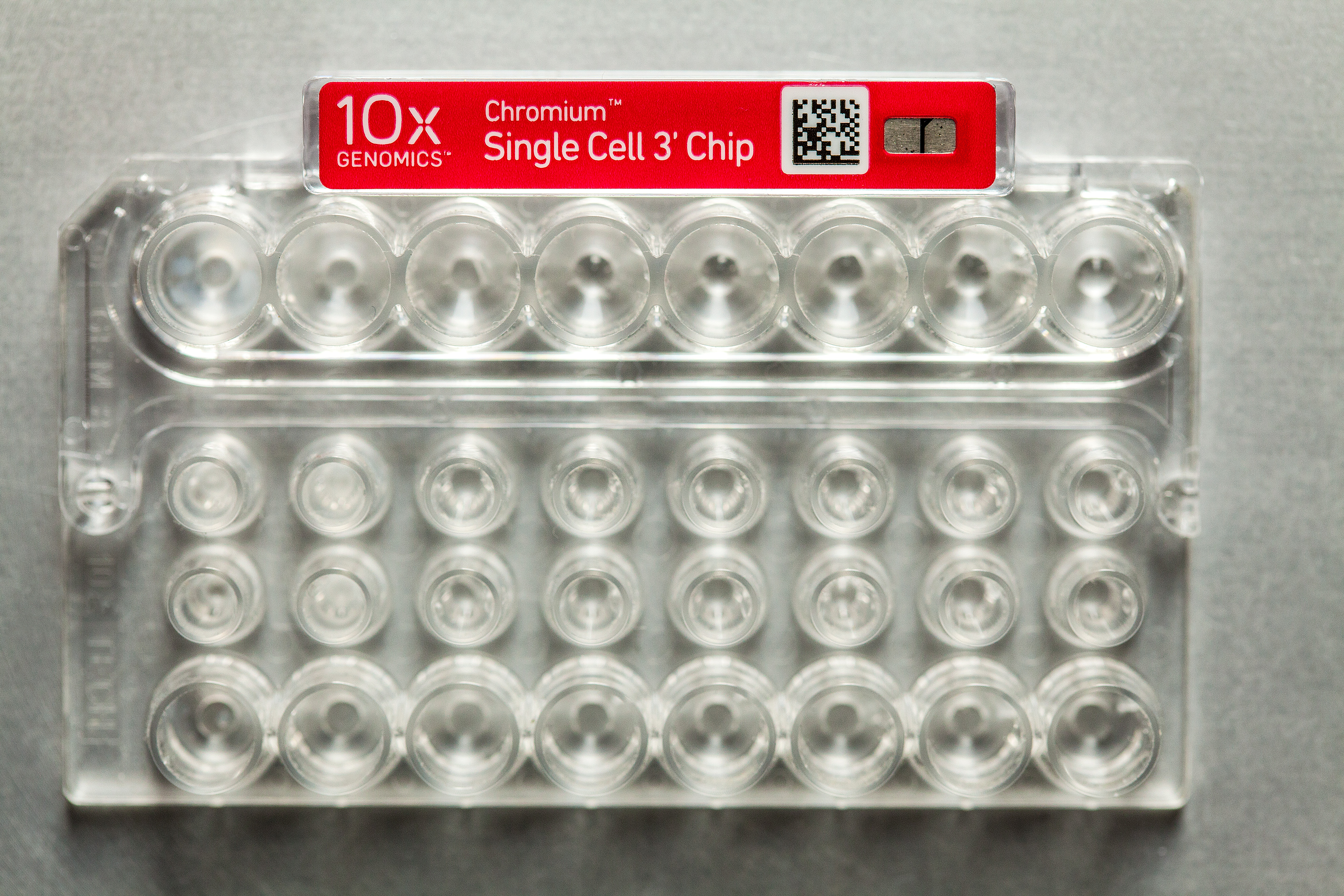
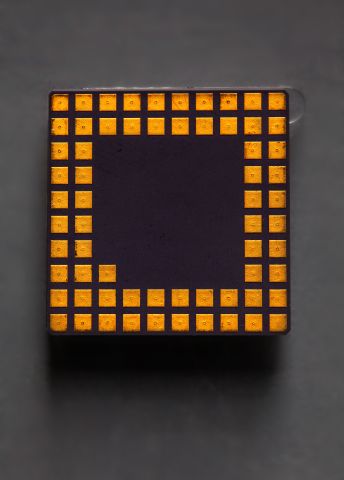
Inconspicuous machine
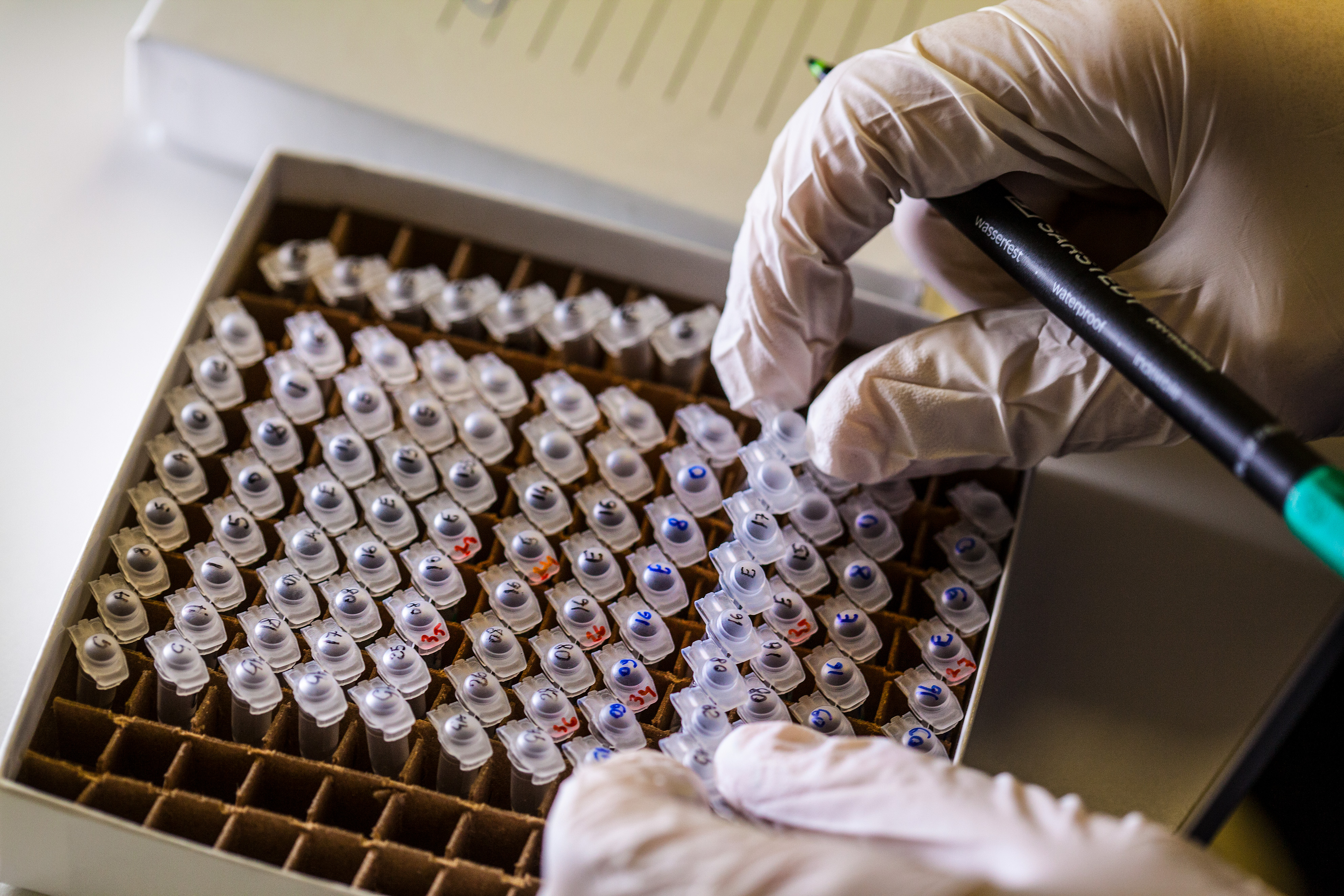
Linnarsson’s team consists of ten people conducting research at Karolinska Institutet, and across the road at SciLifeLab, where the laboratory for single-cell analysis is located. SciLifeLab is a national facility headed by Professor Linnarsson.
The heart of the method is an inconspicuous box standing in the lab. Cells are placed inside, together with a reagent made up of oil and a mixture of enzymes. The machine applies pressure, which forces an emulsion into thin channels. If everything goes as it should, each droplet will contain a single cell. The machine can process 48,000 cells simultaneously. They are then sequenced, making it possible to see the cells in which genes are active.
“Ultimately, it is the DNA sequencing that gives us results,” explains Linnarsson.
Text Carina Dahlberg
Translation Maxwell Arding
Photo Magnus Bergström


