If we better understand the function of proteins in our cells, it will be easier to understand what goes wrong when diseases like cancer strike. Emma Lundberg wants to learn more about what happens when cells divide, and she is happy to enlist the help of computer games to interpret microscope images.
Emma Lundberg
Professor of Cell Biology and Proteomics
Wallenberg Academy Fellow 2016
Institution:
KTH Royal Institute of Technology
Research field:
Mapping cell cycle-dependent proteins
“Our cells are the building bricks of the body, and proteins important building blocks of cells”, Lundberg points out.
Numerous efforts are being made around the world to map the cell and its functions. One such initiative is the cell atlas, which, with Lundberg at the helm, has been developed at SciLifeLab in Solna, north of Stockholm, over the past ten years.
She grew up in the northern Swedish town of Skellefteå, and moved to Stockholm to study chemical engineering at KTH Royal Institute of Technology. But she was more interested in biological applications, so she changed to biotechnology. A year before she gained her PhD she was asked to head the cell atlas project.
“I had done some microscopy work, and this was a chance I couldn’t turn down. Everything we do is aimed at understanding how the human cell works as a biological system. It’s also fun to build something used by others – something that doesn’t exist purely for the purpose of my research. I believe strongly in open science – that research findings should be published openly and accessibly.”
Protein function
The cell atlas forms part of the Human Protein Atlas (HPA), which is funded by the Knut and Alice Wallenberg Foundation. HPA’s goal is to map all human proteins.
“We now know we have approximately 20,000 genes, and they code for various proteins involved in cell processes.”
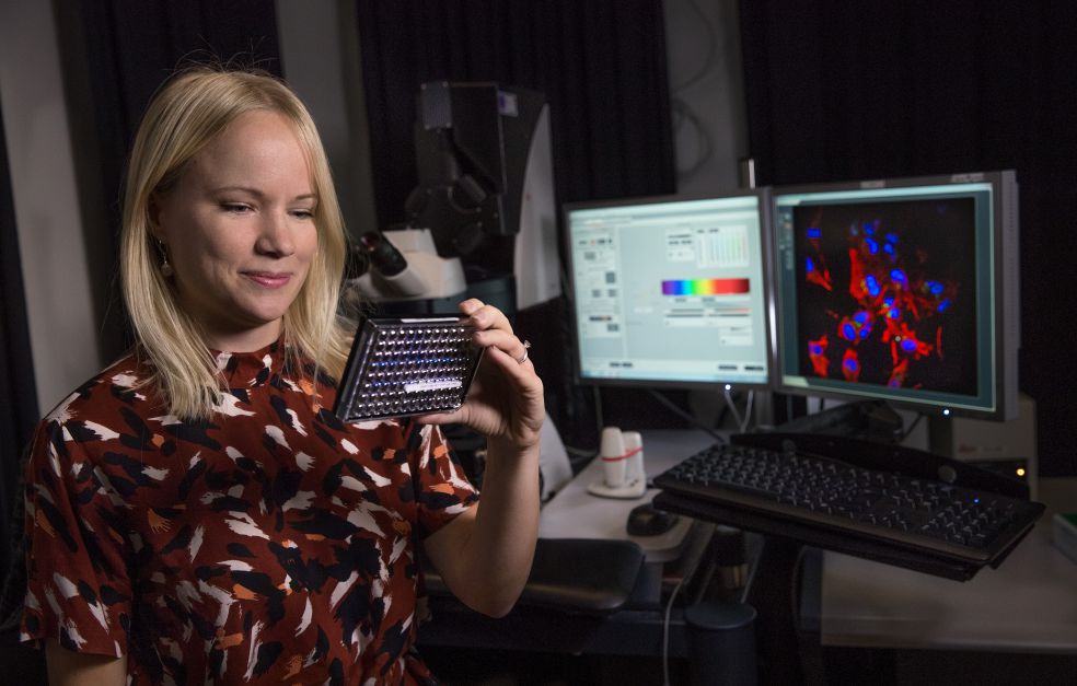
With the help of the 40,000 antibodies identified by HPA, and cells treated with fluorescent dye, Lundberg and her colleagues are able to visualize specific proteins in the cell. The microscope images are published in an online database that receives about 200,000 visits a month.
“We use the cell atlas to map the place in “the little organs” (organelles) of the cell where the proteins are located. This gives us a clue as to their function and context. If, for instance, we know a protein is found in the mitochondria, we can infer that it has something to do with the metabolism.”
Cell cycle in focus
As a Wallenberg Academy Fellow, Lundberg is conducting research into the way that human proteins change as cells divide. The cell division cycle is the basis for life. Shortly after fertilization, the first cell begins to divide – a carefully regulated process that continues throughout our lives.
“The grant means a lot. It gives me the chance to use our cell atlas in a specific and very exciting field to map proteins that vary during the cell cycle. Hopefully, our work will yield benefits in the form of increased knowledge about cell growth and cancer therapies in the future.”
“If the cell division process is poorly regulated, the result may be uncontrolled growth and cancer. There are also other diseases, such as psoriasis, that involve growth problems. We want to understand what happens at molecular level, and find new ways of stopping or slowing down the division process.”
The project is based on the compendious catalog of images in the atlas, and on technology that makes detailed study of individual cells possible.
“It’s really great to be able to transform data from our cell atlas into real knowledge – knowledge that may contribute to the development of new medicines in the future.”
Avatars in computer games
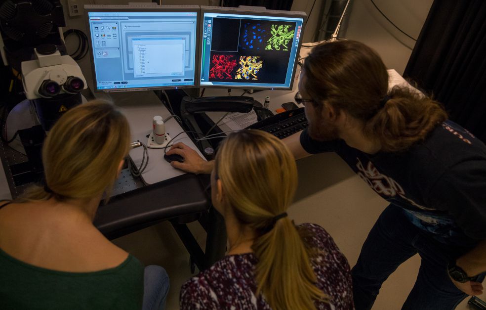
The microscope images are the basis for all work under the cell atlas project. Enormous quantities of data are produced. These data must be interpreted and analyzed. Lundberg explains:
“My project involves advanced image analysis to create a timeline showing how proteins vary with the cell cycle. We also use machine learning, software that has learnt to recognize different phases of cell division.”
They have also received help from the world of computer games. A mini-game called Project Discovery, integrated in a sci-fi game called Eve Online, was developed in collaboration with a young IT entrepreneur from Switzerland and an Icelandic computer games company. The games have helped in classifying patterns in the images.
“After just a couple of weeks we could see that this is a fantastic way of using people’s brains as a resource. Over a 12-month period some 250,000 people helped us to analyze our microscope images, and we obtained 18 million image classifications. These have helped us to find variations in individual cells, and have also helped the project to find new ‘rods and rings’ – a protein structure we know very little about.”
Project Discovery has received very positive feedback from gamers, who appreciated learning more about science and being able to make a contribution. For her part, Lundberg, who lent her identity to an avatar in the game, found that the collaboration whetted her appetite.
“I believe in citizens’ research, and I’m convinced that integration of computer games, AI and open research is the future for medicine and health.”
Cell atlas in the U.S.
Exciting new challenges lie ahead. Emma Lundberg and her family will be moving to the U.S., where she will be working for a year as guest professor at Stanford University, and also at the Chan Zuckerberg Biohub in San Francisco.
“They are producing a cell atlas from a somewhat different perspective. I will continue working on the cell division cycle project, learning more about single-cell analysis, and will be identifying areas in which we can collaborate to understand how the cell works. It’s essential that we are aware of what is going on in the rest of the world so that our work dovetails with that done by others.”
Text Susanne Rosén
Translation Maxwell Arding
Photo Magnus Bergström