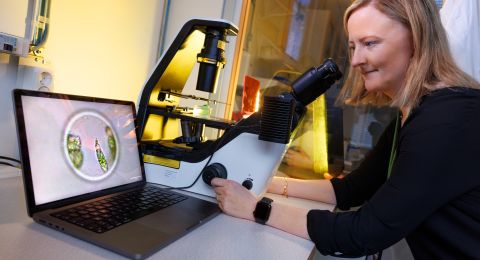
For many years Richard Neutze has dreamed of creating films of the molecular world. His methods have recently seen a breakthrough. Using free-electron lasers, it is possible to capture the minutest secrets of nature on film. This research is contributing to a new biological understanding and may provide the impetus for new medical discoveries.
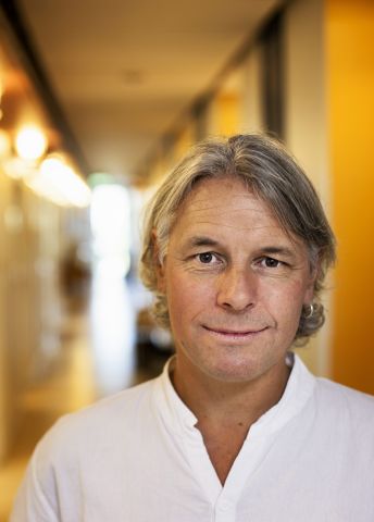
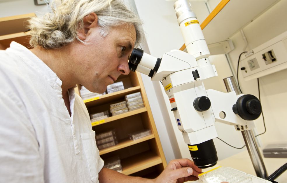
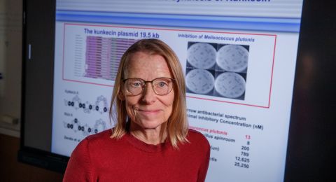
Richard Neutze
Professor of Biochemistry
Wallenberg Scholar
Institution:
University of Gothenburg
Research field:
Structure, dynamics and function in membrane protein structure
The dream was long thought to be science fiction, but is now a reality. Technological developments over the past few years have enabled scientists to produce crystal-clear three-dimensional images of viruses, bacteria and other organisms, and to study their structure in detail. It is now possible to image ultra-fast chemical processes, including photosynthesis, as film sequences.
A new understanding of biology is emerging. Neutze explains:
“It’s a revolution. Now we can film what happens in nature at molecular level in a way we could only dream of twenty years ago.”
The explanation lies in free-electron laser, an instrument that has given research a giant leap forward.
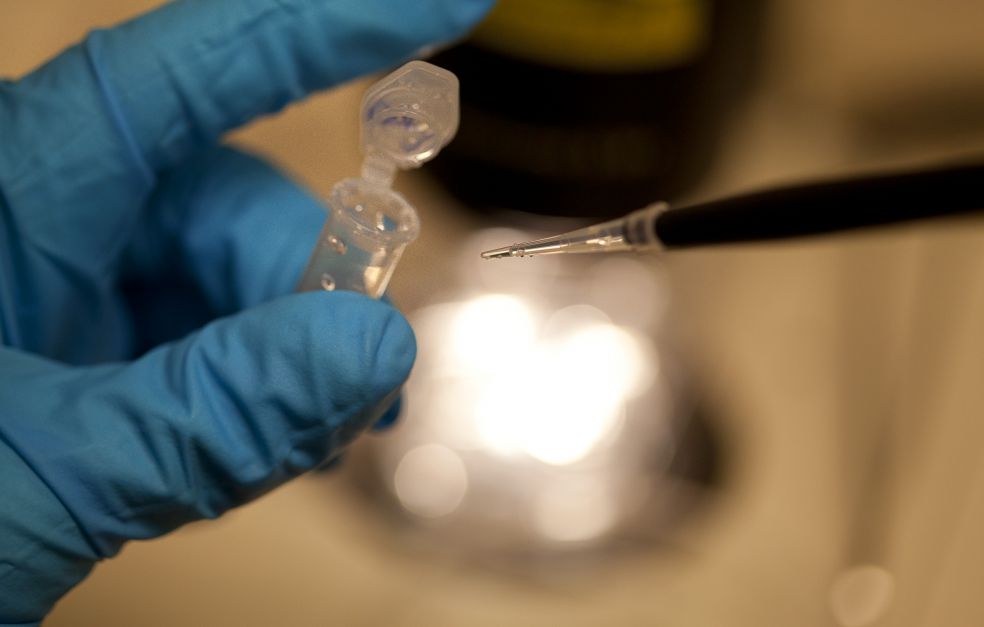
There is actually nothing new about using x-rays to take photographs of proteins. Scientists have been doing so for a long time by directing x-rays through protein crystals. The proteins lie tightly packed inside the crystals in regular patterns, like water in an ice crystal or carbon in a diamond.
When the x-rays pass through the crystal they strike the proteins and are diffracted. Scientists can use the diffraction pattern to produce a 3D image showing the precise atomic structure of the protein.
A billion times more powerful
But the use of free-electron laser completely overshadows the old technology. The free-electron laser shines a beam that is over a billion times more powerful than the x-rays previously available to researchers. Neutze describes the difference thus:
“We used to travel at snail’s pace – now we can move at the speed of light.”
“The Wallenberg Scholar grant was a crucial factor enabling me to continue my research in Sweden. This generous long-term funding has enabled me to build up a competitive team and join the international research environment.”
It should not really be possible to use the laser beam. It is so powerful that it blasts the protein crystals themselves to pieces. But as long ago as the turn of the millennium Neutze and his then mentor, Professor Janos Hajdu at Uppsala University, carried out modeling that showed there to be a time interval that could be exploited. If the pulse was really short – just a few femtoseconds (one millionth of a billionth of a second), they would have time to capture an image of the crystal before it shattered.
Californian facility
It was not until 2009 that these ideas could finally be tried out in practice. The Linac Coherent Light Source – a free-electron laser – had been built at Stanford University in California. It consists of a three-kilometer-long tunnel in which scientists accelerate electrons, which end up being converted into an ultra-high energy x-ray beam. This laser enables Neutze’s research team to follow the movements of proteins at an extraordinary scale and resolution.
One of the first results was the image of a protein involved in African sleeping sickness – trypanosomiasis. It would never have been possible to analyze the minute crystals using traditional technology.
“Science chose the study as one of ten scientific breakthroughs in 2012.”
Capturing ultra-fast protein changes
In recent years the research findings have continued to reverberate around the world thanks to several high-profile publications. In 2018 the Swedish research team had an x-ray film published in Science, showing retinal-based phototrophy, one of the fastest biological processes. Photosynthesis and retinal-based phototrophy constitute the most important energy source for virtually all life on Earth.
The film allows the viewer to see how a retinal molecule is photo-activated with the help of an energy transducing protein. The film shows how the retinal molecule changes shape from straight to curved in the middle, a change that occurs at enormous speed – faster than a millionth of a millionth of a second.
“The change takes about as little time as it takes for light to travel the width of a human hair. It’s amazing that we can see the movement of atoms in a protein on this timescale,” Neutze enthuses.
A similar process takes place in the retina of the human eye. In that case a retinal molecule changes shape under the influence of light, triggering a chain reaction that makes up the mechanism of sight itself.
“The research shows that small, subtle and rapid structural changes are essential for fundamental mechanisms to take place.”
X-ray film of biological photosynthesis
The same methods have also been used to study closely related phenomena. In 2020 Neutze and his colleagues had a study published in Nature in which they managed for the first time to film the short-lived protein changes that occur during photosynthesis.
“That film has given us new insights into how, over billions of years, evolution has optimized light-driven movements of electrons in photosynthesis in order to achieve almost perfect efficiency.”
Neutze reveals that further intriguing films are in the pipeline. One of his aims is to simplify the methods to make them available to more scientists in fields such as medical research and in the development of new drugs.
Twenty years ago Neutze was not sure he would get to see the breakthrough for his research, but the hard work has paid off.
“It’s satisfying to have been part of this research journey – and it’s now the really exciting part begins,” Neutze says.
Text Nils Johan Tjärnlund
Translation Maxwell Arding
Photo Magnus Bergström