As he approaches the end of his research career, Wallenberg Scholar Christer Betsholtz is not resting on his laurels. One of his aims is to provide more knowledge in a heated debate among researchers: how does the brain get rid of waste products?
Christer Betsholtz
Professor of Vascular Biology
Wallenberg Scholar
Institution:
Uppsala University
Research field:
Vascular biology, focusing on how blood vessel cells develop and the part they play in various diseases
The brain is the best protected organ in the body. Underneath the skull the brain is embedded in three membranes that provide protection and structural support. The blood vessels running along the surface and inside the tissue also have special protection: the blood brain barrier. This is made up of special layers of cells in the vessels that stop harmful substances reaching the brain. There is a similar barrier in the middle cerebral membrane, known as the arachnoid mater.
“But unlike the blood-brain barrier, we know very little about how the structure and function of the arachnoid mater. I want to develop new methods and tools to find out more,” says Betsholtz.
The brain’s own waste disposal system
The isolation of the brain from the rest of the body means it has to find its own ways of getting rid of waste products. About half a liter of cerebrospinal fluid – a colorless liquid surrounding the brain and the spinal cord – is produced every 24 hours. This fluid is an important shock absorber, but also contains waste products that the brain needs to get rid of.
“All the metabolites and other waste products produced by the brain must be discarded in some way. But exactly how this is done, and the extent to which cerebrospinal fluid is involved, have led to fairly heated discussions among researchers.”
It is known that more cerebrospinal fluid is produced than is needed for shock absorption. The ventricles surrounding the brain and spinal cord contain only just over 10 centiliters, as compared with the half liter or so produced every 24 hours. The surplus has to drain away somehow, and it is here that the theories diverge. But Betsholtz suspects that the arachnoid mater plays a more important part in drainage than was previously thought.
“Although the arachnoid mater as such has been known since the 1970s, we still don’t understand its importance. But we’ve made some discoveries that may improve our knowledge.”
Barrier of connective tissue cells
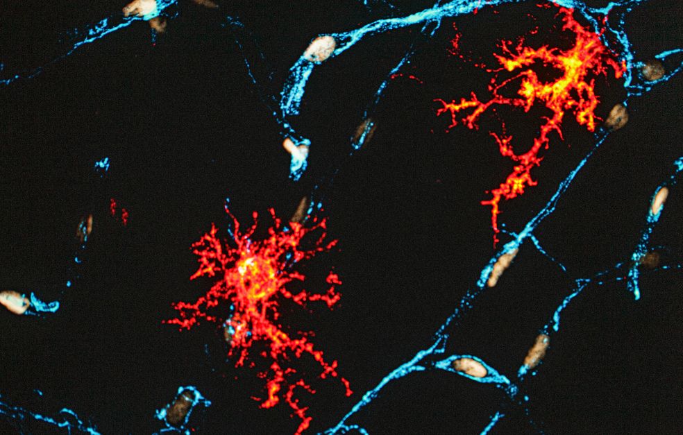
Betsholtz has identified the different cell types in the arachnoid mater using single-cell RNA sequencing. That study revealed that connective tissue cells in the membrane consist of highly specialized fibroblasts, some of which form the barrier itself. In the barrier the cells are packed in extremely thin layers, rather like a stack of pancakes. The layers are also bound tightly to each other to form an impenetrable barrier.
New ways of dissolving the binding, or the “glue,” between the cell layers are needed in order to understand how the barrier works. New tools and methods are also needed to study what happens when the layers are detached from each other.
My research on blood vessel development is a voyage of discovery in the wonderful world of biology.
“We need to know the consequences of destroying the barrier. We place trace substances inside and outside the membrane, enabling us to monitor their progress through the barrier,” he says.
“We know the arachnoid mater heals very quickly when it is damaged during an operation, for example. This probably means it performs a very important function. But what happens while it is healing?”
The first stage of arteriosclerosis
Apart from gaining a better understanding of the function of the arachnoid mater, he wants to delve deep into another research project, aiming to improve our understanding of the first phase of arteriosclerosis. In this disease, fat and substances from blood accumulate on the inside of the blood vessel walls, eventually blocking the blood flow.
“The fat consists of cholesterol particles of a type called LDL. When it reaches into the vessel wall, it causes inflammation. But how does it get past the endothelial barrier? Maintenance and dynamics of the endothelial barrier in the arteries has been very little researched.”
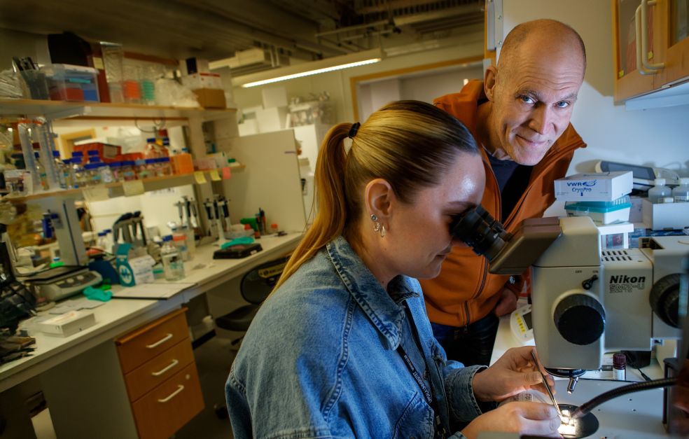
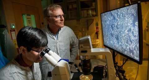
As long ago as 2018 Betsholtz’s research team published the first high-definition molecular map of blood vessel cells in the brain and blood-brain barrier. Since then the mapping process has continued in parallel sub-projects to create maps of organs such as the liver, heart and lungs.
The research team is currently based at two sites: Uppsala University and Karolinska Institutet in Stockholm. Betsholtz himself lives in Gothenburg. He has always divided his time between different cities.
“I used to have an apartment for overnight stays here in Uppsala, but now I live out of a suitcase. I sleep over at friends’ places or with my mother, who enjoys it when I visit.”
He now anticipates a generational shift in the research, in which his role will be to support the research teams taking over ongoing projects. He himself sees no need to wind down, however. On the contrary, the handover gives him time and the opportunity to map out new pathways in what he calls the wonderful world of biology.
“One of the good things about getting older is that you can raise the stakes and take more chances with your research. At my age, you’re not putting your career at risk if you fail. But it has really always been research that has led me rather than me leading research.”
Text Magnus Trogen Pahlén
Translation Maxwell Arding
Photo Magnus Bergström