Step by step, Ilaria Testa has advanced light microscopy to peer inside living cells without harming them. Now the technology is moving on to smart microscopes that can act on their own to capture events of particular interest.
Ilaria Testa
Professor in Applied Physics
Wallenberg Scholar
Institution:
KTH Royal Institute of Technology
Research field:
Development of new microscopy and spectroscopy methods to study the dynamics of life at the nanoscale
Testa has long dreamed of peering into a living cell to study how molecular processes change over time – almost like seeing the bustle of a city from above, preferably with the ability to zoom in on what is happening in each neighborhood.
For a long time this required a compromise between clarity of detail and the risk of damaging the cell due to the powerful illumination required. But the development of super-resolution fluorescence microscopy has changed that.
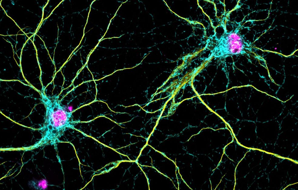
Researchers can now label selected molecules with fluorescent markers and then control which of them should light up, be switched off, or remain dark, enabling them to see details far beyond the limits of a conventional light microscope.
Testa now aims to move on from still images.
“My dream has always been to capture the motion of molecules as films – to study their dynamics using fluorescence and see how they change over time,” she says.
Neurons in focus from the outset
Since moving from Germany to SciLifeLab in Stockholm in 2015, her research team has made a series of major technical advances in microscopy. These have enabled them to push the boundaries of what can be seen in detail – for example, in living brain cells.
“We have studied neurons right from the outset. They are fascinating because so much happens so quickly in a small area. In less than a micrometer, molecules move, signals are fired, and equilibrium is speedily restored. They are perfect systems to study using super-resolution microscopy.”
Her team has published findings showing what happens when neurons communicate with each other. They have also produced three-dimensional images of mitochondria, the powerhouses of the cells, showing that they can contain differing amounts of DNA, depending on where they are located in the cells.
“Our results open up an entire field of new questions, which is perhaps the most interesting aspect of this kind of research. How do synapses get their energy? What drives the rapid processes in these small spaces? Why do mitochondria look different depending on where they are in the neuron?”
Testa has always been keen for her research team to encompass a diversity of competencies. That is why she works alongside physicists, optical engineers, biologists and chemists.
“Early on I was criticized for being too unfocused. But for me it is about balance. We develop systems that other groups should be able to use. Without their perspectives, we risk over-engineering small details that may not have much of a part to play in advancing the field,” she explains.
Smart microscopes
The next step is to combine these technical advances into a microscope that can independently capture events of particular interest in a cell sample – changes that may be over in a microsecond.
She has likened it to trying to see a shooting star on a dark autumn night: for a human it requires concentration, patience and a good measure of luck. But for a machine it is different.
My dream has always been to capture the motion of molecules as films – to study their dynamics using fluorescence and see how they change over time
The microscope should have a drone-like view of everything that happens and the ability to zoom in at lightning speed to show details at the highest possible resolution.
Above all, it should be able to anticipate a change before it happens.
“The microscope will react using sensors in neurons that light up when something changes, such as calcium concentration or pH level. Its speed will enable us to capture events that previously could not be observed.”
The microscope should also be able to capture three-dimensional movements in the cell, such as the rotation of molecules as they change or merge.
Extensive software development is required to handle the complex technology. AI-based image recognition will give the microscope the ability to detect shape changes, and artificial intelligence will also help adapt the programming to different environments. The ultimate goal is for the entire system to be as open as possible to achieve maximum reach.
“We want all the tools we develop to be open and distributed via open source. Other researchers should then be able to make their own additions, for instance by adapting the instrument to other tissues.”
Captivated by the visual
Testa’s research career began with an interest in mathematics when she was growing up in Genoa, Italy.
“In high school I had a fantastic mathematics teacher: she was inspiring and brought mathematics to life. Her husband was a physicist and shared insights into theoretical physics.”
Testa has always had a parallel interest in art and culture. Every conference trip includes a museum visit. Her leisure interests include photography and painting.
“I’ve always been drawn to images and form. When I saw the first pictures from my experiments, I was captivated both by the visuals and by the mysteries of why an image looks the way it does. Perhaps that’s why I find so much inspiration in microscopy: it makes me feel a bit like an artist.”
Text Magnus Trogen Pahlén
Translation Maxwell Arding
Photo Magnus Bergström