To be able to run away from danger or creep up on prey, it is essential that our muscles perform the right movement, at the right speed, at the right time. With the help of zebrafish, researchers at Karolinska Institutet are now delving deep into the neural processes involved in the control of movement. In the longer term, the research may contribute to new therapies for spinal injuries and neuromuscular diseases.
Project Grant 2018
Decoding the logic of the neural circuits for motor actions
Principal investigator:
Abdel El Manira, Professor of Neuroscience
Institution:
Karolinska Institutet
Grant in SEK:
SEK 16.2 million over five years
Human behavior is based on movement. The way we move has to be constantly adapted to the situation, whether running to catch a bus or making our way quietly around a library.
Movements are performed by our muscles, which are controlled by signals from the brain. The signals reach the muscles via neurons in the spinal cord. A complex exchange of information in the networks of neurons in the brain and spinal cord is needed before the signal to perform a movement is sent from the brain to the muscles. The structure of these networks is largely an unsolved biological puzzle, now being examined in a project funded by Knut and Alice Wallenberg Foundation.
“We're trying to understand how the brain converts thoughts into actions – how we process information from our surroundings to perform a movement adapted to a given situation,” says Abdel El Manira, Professor of Neuroscience at Karolinska Institutet, and lead researcher.
Aided by aquarium fish
Most of the research is being performed on zebrafish. These centimeter-long aquarium fish are popular in neurobiological research, partly because they are amenable to genetic modifications. The researchers in the project are studying adult fish, whose muscles and neurons are fully developed. The experiments are therefore being performed on fish that are six to twelve weeks old.
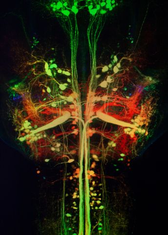
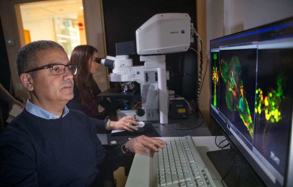
On a computer screen connected up to a microscope, El Manira shows an image of a zebrafish brain. The structures are shown in fluorescent colors. The 300–400 neurons that run from the brain to the spinal cord are visible in neon green.
“Isn’t it amazing that the brain can control so many behaviors with so few neurons? And it’s fascinating that they can be so readily studied. Here we can see neurons that control escape behavior – these are for rapid movements, while others control slow movements. Our experiments have revealed these differences.”
The researchers hope that the knowledge they gain with the help of zebrafish can be transferred to other animal species that have similar muscle structures – humans for example. But the nervous systems of humans and other animals are more complex, and not as easily analyzed in detail as that of zebrafish.
Measuring individual neurons
One method the team is using is called optogenetics. This involves modifying neurons with a gene that produces a light-sensitive protein. They can then be turned on and off using a light source, enabling the researchers to stimulate specific neurons in the brain and spinal cord to see which muscles they control.
The researchers also attach electrodes to individual neurons, and stimulate them to figure out how the cells are connected to each other. They also remove specific neurons to see how this affects fish behavior, e.g. how they swim, and to find out how many neurons of a given type are needed for the action.
“The grant we have received has enabled us to broaden our technical platforms. Now we can tackle more conceptual questions that remain unresolved, instead of merely carrying out the most obvious experiments.”
Progress made so far by the researchers includes the insight that zebrafish muscles are not all controlled by a single network; there are three separate networks controlling the slow, medium-speed and fast muscles respectively.
“Instead of activating all neurons simultaneously for all behaviors, only the parts that are needed are activated. When the first network has reached its maximum speed, the next one kicks in, like a car shifting up a gear to achieve higher speeds.”
Recently, El Manira’s research team demonstrated that the neurons activating muscle cells, known as motor neurons, are more than mere passive channels for information from the brain. They can also carry information back to the brain. The findings were published in Nature, and are now being further examined as part of the project. The researchers are trying to learn whether the muscles send back signals that impact the organization of the neural network.
Knowledge about movement and disease
The project aims to generate new fundamental knowledge about the structure of the networks controlling our movements. Insights into normal function will also increase our understanding of what happens when the nervous system is damaged. El Manira elaborates:
“Our findings offer potential for further research into spinal injuries and motor neuron diseases such as ALS. They may also contribute to the development of new therapies for restoring motor function impaired by those diseases.”
The project runs for five years. At the end of that time El Manira thinks they will have gained a fairly good idea of how the networks in the spinal cord are interconnected to control speed of movement and escape behavior, and which parts of the brain control swimming at different speeds and escape.
“I also hope that we will have begun to find out whether these networks are similarly organized in mammals. We’ve made rapid progress so far, so it ought to be possible,” he says.
Text Sara Nilsson
Translation Maxwell Arding
Photo Magnus Bergström