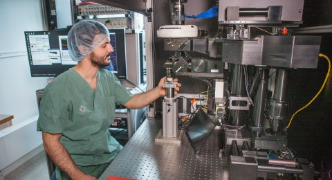
Joakim Lundeberg's laboratory will study the brain, cell by cell. The research team has developed a pioneering method for studying what genes are active in individual cells. In a single experiment, they can study more than two million cells in a tissue. The project will generate a unique map over every part of the brain. First out: the olfactory center.
Project Grants 2012
Spatial transcriptomics of the brain
Principal investigator:
Joakim Lundeberg, Professor of Molecular Biotechnology
Co-investigator:
Jonas Frisén, Karolinska Institutet
Institution:
KTH Royal Institute of Technology
Grant in SEK:
SEK 16.7 million for a two-year project with the possibility of seeking a further three-year extension.
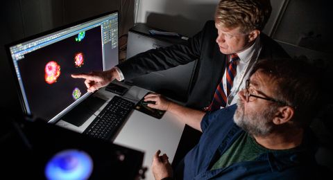
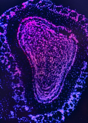
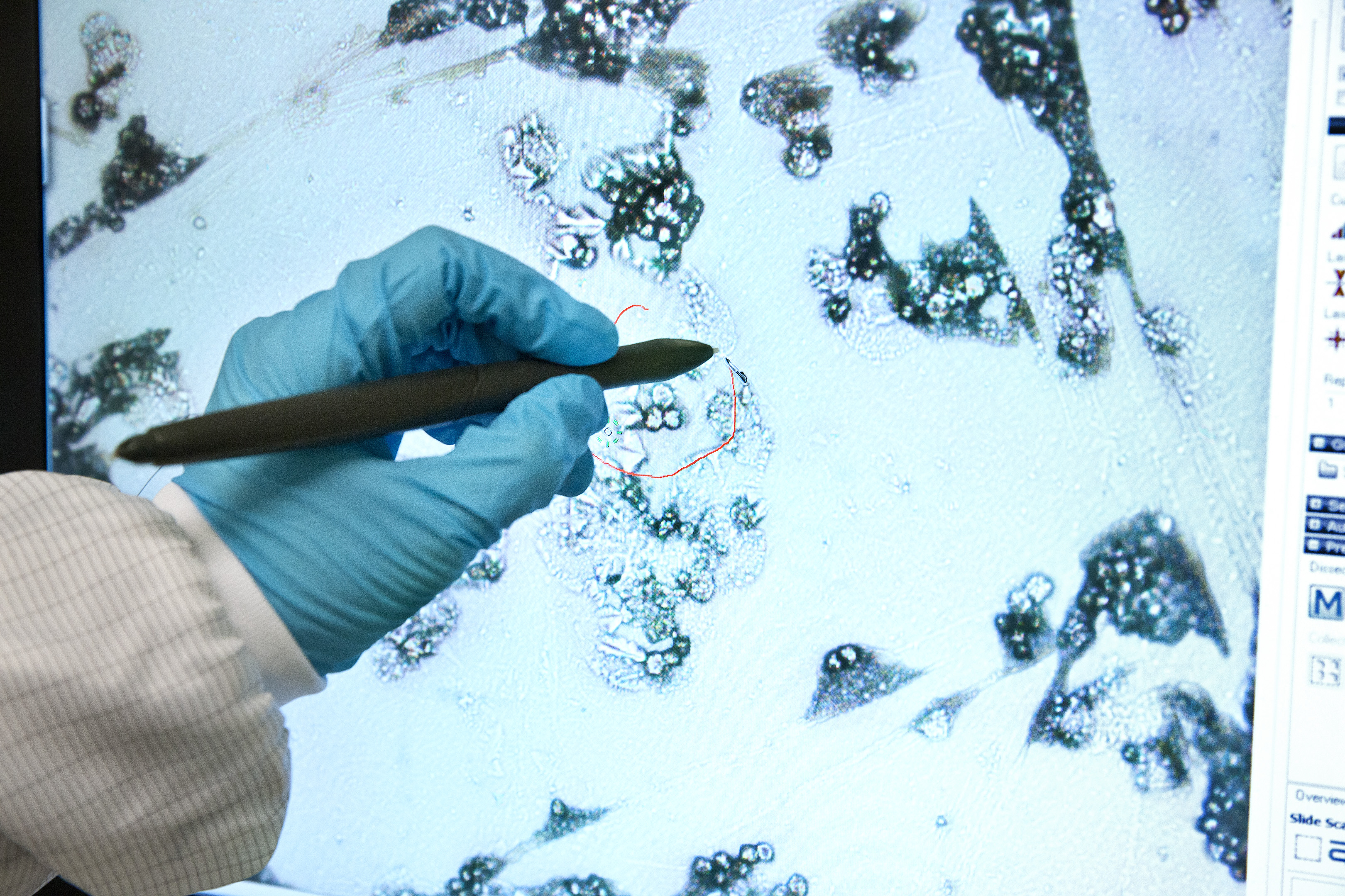
“It has such an attractive morphology. Its shape looks like Africa,” says Joakim Lundeberg, Professor of Molecular Biotechnology at the SciLifeLab in Stockholm.
He points to a picture of the olfactory center that he clicked up on the computer screen. All receptors that allow us to sense smell are in the olfactory center. The tissue section that is on the microscope glass is extremely thin. Joakim Lundeberg can, if he wants, zoom in on every single little cell.
When he clicks further in his slide presentation, a grid is superimposed on the olfactory center; a white grid where every box is 13x13 micrometers. One cell is approximately 10 micrometers in diameter. So there is space for around one cell per box. Joakim Lundeberg can choose which box he wants so the computer can tell him exactly what genes are active in the cell that is in the box.
When he talks, it all sounds so simple. However, the technology he has developed is ingenious: never before have researchers been able to map the body on such a detailed level.
Revolutionary innovations
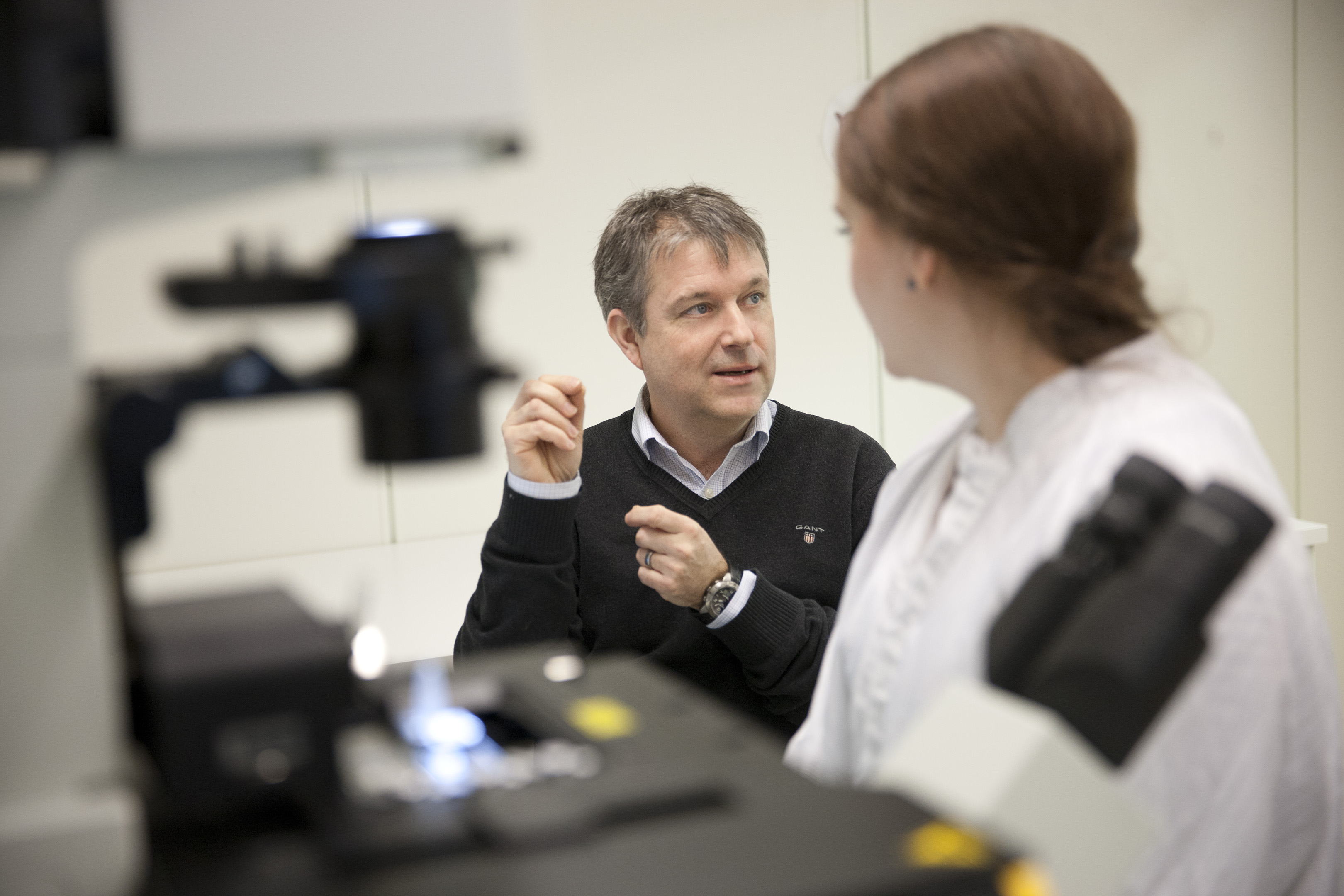
The technology that Joakim Lundeberg, together with Professor Jonas Frisén and post-doc Patrik Ståhl, is now developing in the Wallenberg-financed project “Spatial transcriptomics of the brain” will probably revolutionize the view of the body's tissues. In order to understand how a tissue works, researchers need to know what genes are active. Researchers previously cleaned out cells from the tissue and put them together. They looked at that cells in clumps and found out how they function - on average.
What is unique about the method is that the researchers can study the gene activity in every individual cell in a piece of tissue. They will therefore be able to understand in a completely new way how cells cooperate to form a whole. To begin with, they will study our thinking organ:
“We will map the genes that are active in the brain in a very large-scale manner. We will describe all cells in the brain based on their gene activity,” says Joakim Lundeberg.
Traditional pathology is combined with modern biotechnology
He depicts the technology his research group is developing as a coalescence of traditional pathology, where doctors study thin sections of a tissue in a microscope, and modern biotechnology where a chip can be used for large-scale mapping of genes. They coat the microscope glass with something that can be compared with molecular biology fishing rods. These consist of RNA molecules, a chemical cousin of the DNA molecule that builds our genes.
When a gene is active in a cell, a copy of the gene is created in RNA. This copy is called messenger RNA. The molecular biology fishing rods in the microscope glass are designed so that they can capture all messenger RNA in a cell. In addition, all fishing rods that are in boxes of 13x13 micrometers differ from all other fishing rods on the microscope glass. Every box has its own address tag. This means that the researchers can sort out all material in their gene analyses. They know which messenger RNA come from which part of the microscope glass. When they look at the microscope picture, they can see exactly which cell was in that box. They thereby know exactly which genes are active in different cells.
“This chip is five by five millimeters and is divided up into 135,000 different small boxes. This way, we can analyze 135,000 cells at a time. But if we can produce a chip with two million boxes there, we can look at two million cells in an experiment,” says Joakim Lundeberg when he points at the Africa-shaped olfactory center.
Will revolutionize the view of the body
His research team will now study all of the brain's key parts and create an atlas over the gene expression of cells. They will take up the competition with a similar project that is under way in the US where the researchers are using a more unwieldy method.
“They are looking at one gene at a time and need to do 20,000 experiments for every part of the brain. It takes a huge amount of time,” says Joakim Lundeberg.
The US project has a budget of USD 500 million. The research team at the SciLifeLab will do the same thing significantly faster for a fraction of the cost. And when the actual atlas is in place, they will begin studying various diseases, such as Parkinson's, cancer and schizophrenia. The method can also be used on other tissues in the body. It is an entirely new tool that will generate knowledge that researchers were previously only able to dream of.
Text Ann Fernholm
Translation Semantix
Photo Magnus Bergström