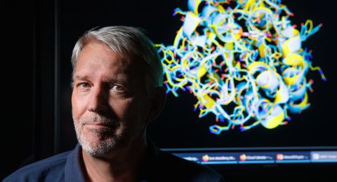
Research into proteins – the building blocks of life – is now taking a giant leap forward. New methods enable scientists to image proteins in 3-D, and in more detail than ever before. This paves the way for better drugs and new fundamental discoveries in the biochemical field – and means Xiaodong Zou has realized her dream.
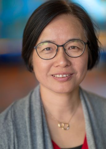
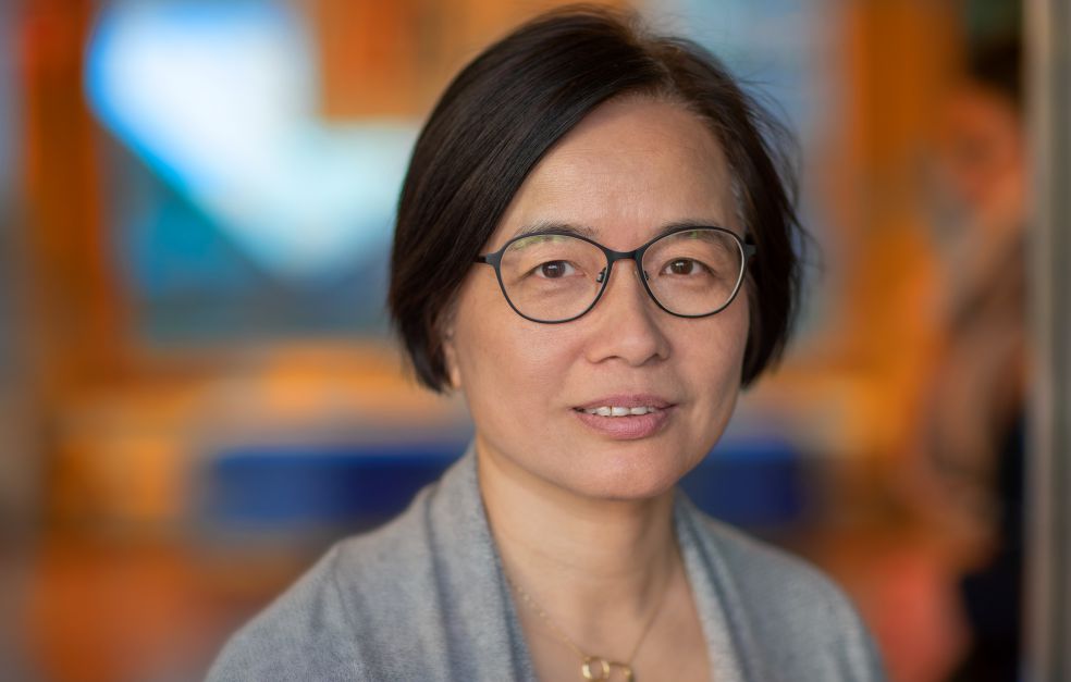
Xiaodong Zou
Professor of Structural Chemistry
Wallenberg Scholar
Institution:
Stockholm University
Research field:
Structural chemistry and electron crystallography. Electron crystallographic methods of studying the atomic structure of a material or molecule.
The idea of imaging the structure of proteins was born when Zou was a young PhD student. She realized the importance of exploring biological molecules, particularly proteins – the molecules that do most of the work in all living systems, from bacteria to plants and humans. Without knowledge of the structure of molecules, we can only make guesses about their characteristics. But the technology was not available at the time, and her plans had to be shelved.
“I tried to make protein crystals even then, but didn’t succeed, and my supervisor said the project was too difficult. But I never gave up the idea,” Zou recalls.
For many years she instead focused on inorganic materials, particularly porous materials, and worked tirelessly to develop the technology. Among other things, she led a project funded by Knut and Alice Wallenberg Foundation that made it possible to produce high-resolution three-dimensional images of materials using an electron microscope.
“The support from Knut and Alice Wallenberg Foundation has been crucial to my career in Sweden. I was about to move to Oslo when I was awarded one of the seven Royal Swedish Academy of Sciences special research positions in 2000, with a grant from Knut and Alice Wallenberg Foundation. Being chosen as a Wallenberg Scholar means I can now concentrate my research on important and difficult aspects of electron crystallography.”
A doorway opened to a new world at nanoscale, in which the researchers can see how the atoms that form the material are arranged. This in turn enables them to tailor-make materials with desired properties. The vision is that the same technique will also be of benefit when it comes to the human body.
“There are so many interesting questions that can be examined in a new way, such as how different biological systems work, and how more effective drugs can be developed to combat diseases,” Zou says.
World-leading research
Thanks to the Wallenberg Scholar grant, she can now bring her dream to life and derive benefit from the experience of many years spent developing electron microscopy. Her research team at Stockholm University is the first in the world to study previously unknown proteins, and has published several sets of findings that have attracted wide attention. The aim is that Sweden should become world-leading in this field.
“We have gone from nothing to being one of the world’s leading research teams in structure determination of protein crystals using electron diffraction.”
Different methods have different limitations. The most widely-used method used to be X-ray diffraction, which requires access to costly synchrotron facilities. To perform analyses, researchers are obliged to reform the substance in question into fairly large crystals, which is a challenge with biological macromolecules such as proteins.
Fantastic new images
Electron diffraction is a quicker and more readily available technique.
“It’s easier to build a facility “in house”, which means that more research teams can benefit from the method, as everything is under the same roof.”
Scientists can now produce increasingly detailed high-resolution images at a faster pace than before.
“Electron diffraction enables us to obtain fantastic images of crystals smaller than one micrometer. This can be compared with X-ray diffraction, which needs crystals that are tens of micrometers across. The reason is that the electrons interact much more strongly with the atoms in the crystal than is the case with X-ray diffraction,” Zou explains.
Better drugs
Zou is now researching using a variant of the method called MicroED. It is used to study protein microcrystals. One advantage of the method is that MicroED can also provide information on atomic charge. This yields completely new insights into biochemical processes, since the proteins involved in the reactions often assume different charge states as their atoms gain or lose electrons.
MicroED may pave the way for better drugs, for instance in a field known as “fragment-based drug discovery”. This entails a series of structure determinations of hundreds of protein crystals, to ascertain the fragments to which a protein binds, and the location of the binding points in the protein. This information can be used to design new drug molecules. MicroED can also be used to find out how a drug molecule interacts with the protein.
“We also want to develop increasingly automated analyses,” Zou says.
Images of protein molecules using cryogenic technology
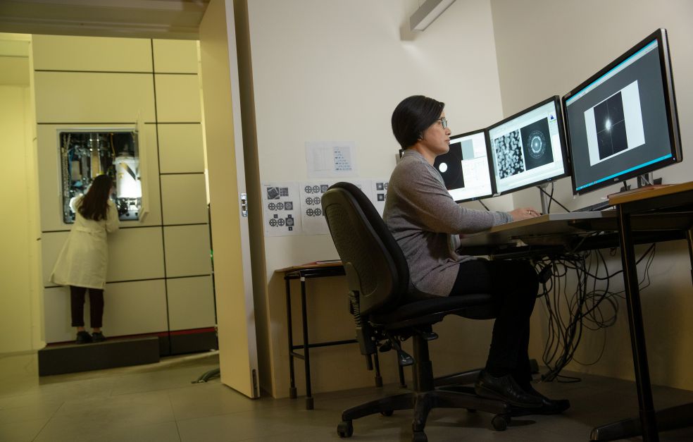
To explore relatively large protein molecules, Zou is using cryogenic electron microscopy, a Nobel Prize-winning method, in which the object to be analyzed is embedded in a thin layer of ice. The method has been refined so that it can also be used on protein microcrystals. The solution containing protein crystals is plunge-frozen to minus 170 degrees Celsius using liquid ethane. The sample is then analyzed under the cryogenic electron microscope. This involves placing a beam of electrons at microcrystals to produce images of protein molecules.
Zou is continuing her work using the entire arsenal of methods at her disposal, and wants to optimize everything – from microscopy and software to data collection and analysis. The idea is to establish cryogenic electron microscopy as a leading method for structure determination, and to ensure that it becomes more widely used.
“We have noticed a great interest among researchers and companies, and want to do whatever it takes to exchange ideas, train students and establish this field in the international research arena.”
Text Nils Johan Tjärnlund
Translation Maxwell Arding
Photo Magnus Bergström