New technology is enabling scientists to better explain the interaction between sperm and egg in mammals. Cryo-electron microscopy is being used to gain a more detailed picture of fertilization. The research has already aided our understanding of infertility.
Project Grant 2018
Integrative structural biology of mammalian fertilization: Unveiling the beginning of life from gametes to atoms
Principal investigator:
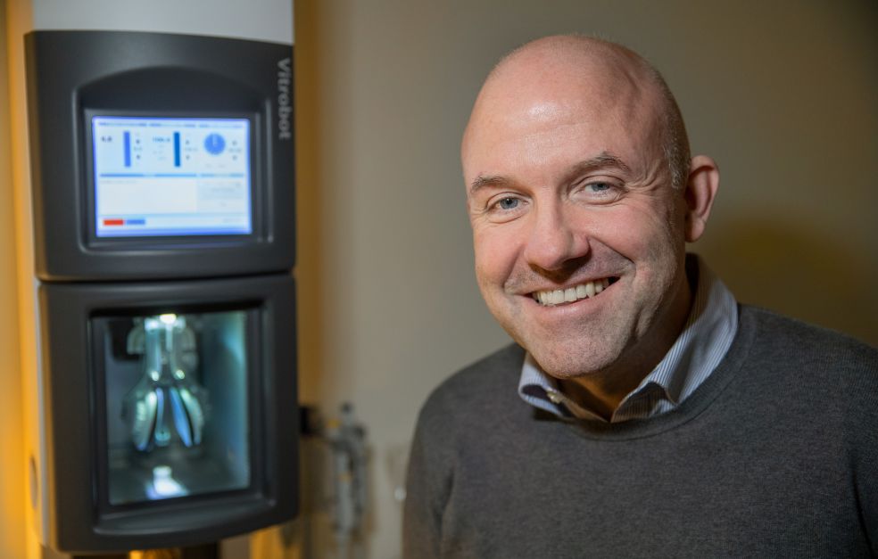
Professor Luca Jovine
Co-investigators:
SciLifeLab
Alexey Amunts
Umeå University
Linda Sandblad
Institution:
Karolinska Institutet
Grant in SEK:
SEK 29,400,000 over five years
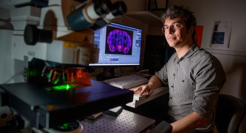
Lennart Nilsson, a scientist and photographer, may well be the person who did most to spread knowledge of how a baby is conceived using the images he captured with a scanning electron microscope. The photographs were taken in the 1960s, and have been a source of inspiration to Luca Jovine since he was a child. Now Jovine is heading a new project whose goal is to dramatically zoom in on this event by showing what happens at molecular level when sperm and egg meet.
“After first approaching this biological problem with X-ray crystallography, we will now further visualize fertilization with the help of cryo-electron microscopy. If we can bridge the knowledge gap between cell and molecular level, we will get closer to solving the riddle of fertilization,” says Jovine, who is a professor of structural biology.
Multiple stages of fertilization
The research project follows the sperm’s journey from its first contact with the outer coat of the egg cell to the point at which it merges with the egg plasma membrane. The researchers are adopting a twin-pronged approach to their task. One goal is to better understand how the sperm and the egg coat recognize each other at the onset of fertilization. The second goal is to improve our understanding of what happens when the two sex cells (gametes) fuse to form the first cell of a new individual – the zygote.
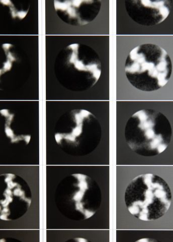
Jovine’s research team is among the world leaders in the field. As long ago as 2010 they were the first to map the complete three-dimensional structure of ZP3 – a protein component of the vertebrate egg coat to which sperm binds.
“We’re the first research team in the world that has adopted a systematic approach to understanding fertilization with the aid of structural biology,” he says.
In 2017 they were also able to reveal how a sperm protein binds to an egg coat protein at the atomic level, a contact that initiates the fertilization process itself. As part of that study, they also compared two egg coat proteins, one from a mollusk and the other from a human. The results were very surprising: despite being very different in sequence, the VERL mollusk protein was found to share the same three-dimensional structure as its human equivalent – ZP2.
“It was an astonishing discovery, given that 600 million years of evolution separates mammals from mollusks. But we still do not fully understand what are the implications of this structural similarity for the binding process itself.”
New technology opens the door
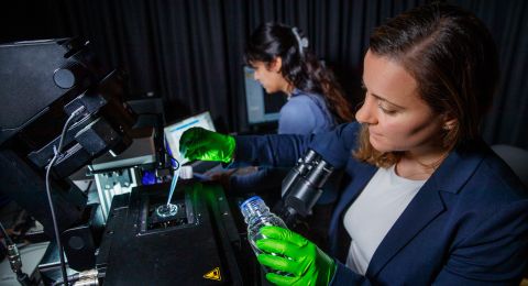
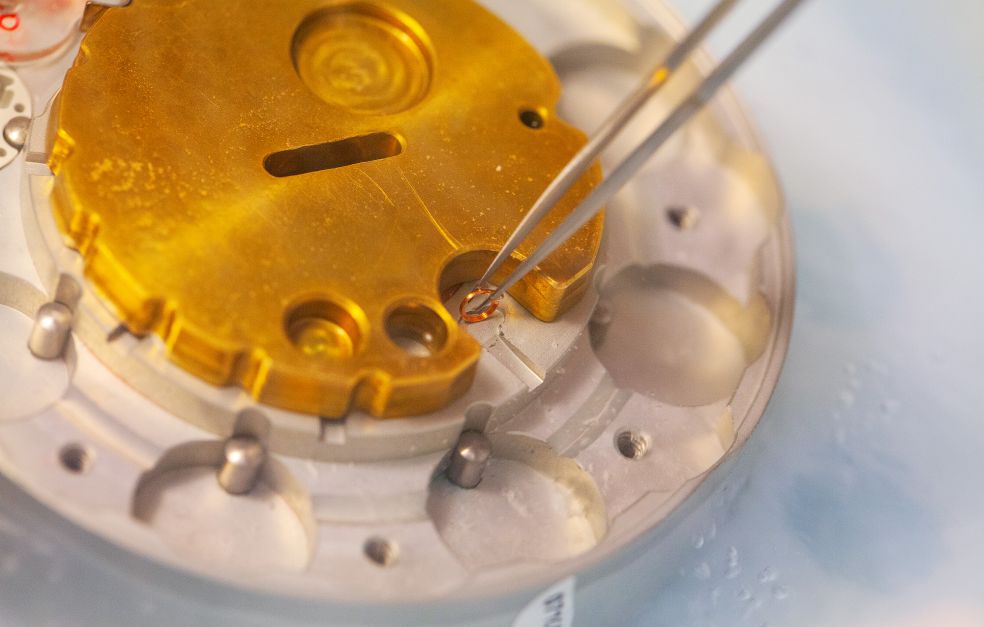
Although the research team has so far largely used X-ray crystallography, the new project will start employing several forms of cryo-electron microscopy, combined with more traditional biochemical and cellular methods.
“By combining different techniques, we hope to be able to ultimately show all levels, from high-resolution visualizations to what can be seen under a normal microscope.”
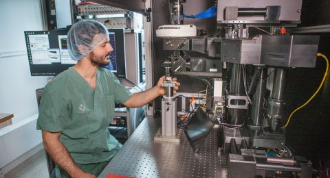
He believes that technological developments have brought the fields of structural biology and cell biology closer together. This is why access to SciLifeLab is key to further progress. A further success factor is collaboration with the team of Alexey Amunts, an expert in cryo-electron microscopy.
“There aren’t many places in the world that combine the state-of-the-art equipment needed with the right knowledge in this way,” Jovine explains.
The project also involves Sweden’s other national cryo-electron microscopy node: Umeå Core Facility Electron Microscopy (UCEM). Microscopist Linda Sandblad, who is director of facility, is also involved in the project.
The next stage: fusion
Once the sperm has penetrated the egg coat, it binds to the plasma membrane of the egg cell and the two gametes fuse. Two of the key proteins in the fusion process, Juno and Izumo1, have been structurally characterized by Jovine’s team as well as other research groups. But it still remains to ascertain what actually causes fusion to occur.
“We want to understand the phenomenon by visualizing it. But it’s probably a very short process, which makes it more difficult to capture.”
A further complicating factor is the size of the egg cell itself.
“The egg cell is the body’s largest cell, and there are a number of technical hurdles we must overcome when we prepare it for analysis. It is a little easier to study how the sperm binds to the egg coat, since the latter can be removed from the egg cell.”
New insights into infertility
The research leading up to this project has already had an impact in the health care sector. Jovine explains:
“We have seen how doctors used our findings to gain a better understanding of female infertility. Using individual gene sequencing they have been able to detect mutations in the same egg coat proteins whose structure we determined, and started making use of this information to explain the patients’ infertility.”
Although the problem of infertility can often be surmounted with the help of in vitro fertilization, studies in mice suggest that mechanical fertilization of this kind may give rise to other complications.
“No one yet knows whether this also applies to humans. It would naturally be best if we could find ways to aid the natural fertilization process. This is highly complicated and involves selection processes that we still don’t understand.”
The new research project may also open the way to develop new forms of hormone-free contraceptives. The proteins involved in fertilization can serve as targets for completely new kinds of agents interfering with this process.
“We don’t know yet what we’re going to discover – and that is precisely what makes the project so exciting,” Jovine concludes.
Text Magnus Trogen Pahlén
Translation Maxwell Arding
Photo Magnus Bergström