An entirely new way of studying the innermost core of life has opened up. Thanks to the development of x-ray lasers, it will soon be possible to take razor-sharp three-dimensional pictures of virus particles, individual cells and live bacteria at an atomic level. Something that can be of major scientific significance.
Project Grant 2011
Photon science and x-ray lasers
Principal investigator:
Janos Hajdu
Co-investigators:
Uppsala University
Joseph Nordgren
Raimund Faifel
Swedish University of Agricultural Sciences
Inger Andersson
Institution:
Uppsala University
Grant in SEK:
SEK 36.5 million
Biologists have long nurtured a dream of depicting individual biological objects without having to cut, freeze or contrast dye them with metals, which is necessary in electron microscopy. The wish has been to be able to take high-resolution pictures of viruses, organisms and bacteria, and on a detailed level capture how these tiny structures are built. Something, which in turn can provide unique knowledge on how viruses are spread and take hold, about disease development and evolutionary connections and provide inspiration for new medicines.
Seen as science fiction
But this hope has long been seen as science fiction, explains Janos Hajdu, who is a driver in the field and Professor of Molecular Biophysics at Uppsala University. His interest was incited in 1994 in the synchrotron lab in the village of Daresbury outside Liverpool.
“The idea was then born of using free electron lasers and extremely intensive, short x-ray pulses to make depictions of the biological material before it is destroyed by the laser beam. We discovered that there was a time interval before the destruction that we could utilize.”
Little Daresbury with a cluster of houses, a church, a vicarage and a pub is the birthplace of author Lewis Carroll, known for “Alice in Wonderland”. And a wonderland was exactly what the research could lead to, if the theories proved to be correct. But at the time, they were dismissed as unrealistic.
“We had to work on this in our free time. It was not a main project, but the thoughts on it often disrupted the night’s sleep. The challenge was calculating if there was enough time to take pictures before the molecule was destroyed. Through simulations in computer models, we gradually realized that, yes, we would indeed have time!”
The biological samples were exposed to extreme heat from the x-ray laser, 200,000 degrees Celsius, and were explosively destroyed when struck by the beam. The concept is based on the ultrafast x-ray flash having time to yield a diffraction pattern before the explosion. This pattern in turn makes it possible to recreate the image of the biological sample.
World’s first free electron laser
Janos Hajdu also found love in Daresbury, which led him to Uppsala in 1996. There, he and his partner Inger Andersson, who is Professor of Plant Biochemistry at the Swedish University of Agricultural Sciences, conducted the research and contributed to building up the world’s first free electron laser facility, placed at Stanford University in California.
With the help of this facility, the Uppsala researchers have now been able to demonstrate that the principle works in reality. This has resulted in the first three-dimensional image of a virus, mimivirus, which previously could not be studied in such detail. With a conventional method like electron microscopy, the possibility of an in-depth picture was very limited due to the virus’ size and special surface structure.
The mimivirus has an inner-structure reminiscent of a cell nucleus, and there is speculation that this kind of virus and eukaryotic cells are biologically linked.
“The mimivirus codes, for example, for more protein than what it needs, and bears a large backpack with genes that it apparently does not use, a sign that there is a connection we are not aware of. There is a blind spot on the inside of life, and it is there that we now hope to be able to fill in information thanks to these detailed images,” says Inger Andersson.
Even the influenza virus H5N1, HIV and Ebola could be studied with the x-ray laser in the future, and the new knowledge that comes forth may contribute to greater understanding of a number of diseases. If, for example, the mechanism for how a virus infects its host is known, it may also be possible to stop its progress, says Inger Andersson.
Pictures of photosynthesis
The research team also hopes to be able to take pictures of the basic processes in photosynthesis. A step in the right direction, is the recent depiction of cyanobacteria, which live on photosynthesis and are found in water worldwide, including Sweden. The unique pictures show the atomic structure of protein complexes in the cell membrane that capture sunlight and convert it to energy in the bacteria.
Membrane proteins are important in various living processes, not just as energy converters, but also as the cell’s transporters and as receptors for pharmaceuticals. The new depictions can therefore revolutionize the biological research.
Now, Janos Hajdu is also involved in the building of the first free electron laser in Europe, XFEL in Hamburg, which is paving the way for further experiments. The grant from the Knut and Alice Wallenberg Foundation makes it possible to double the research team in Uppsala from 15 to 30 people and recruit prominent names.
“We’ve learned through scientific history to be prepared for impasses. But now, the critics have fallen silent and many are instead following in our footsteps. It’s a fantastic feeling that we can now study the living cell. In the next five years, we will make many exciting discoveries,” says Janos Hajdu.
Text Nils Johan Tjärnlund
Translation Semantix
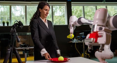
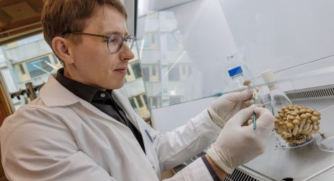
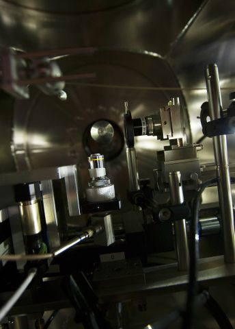
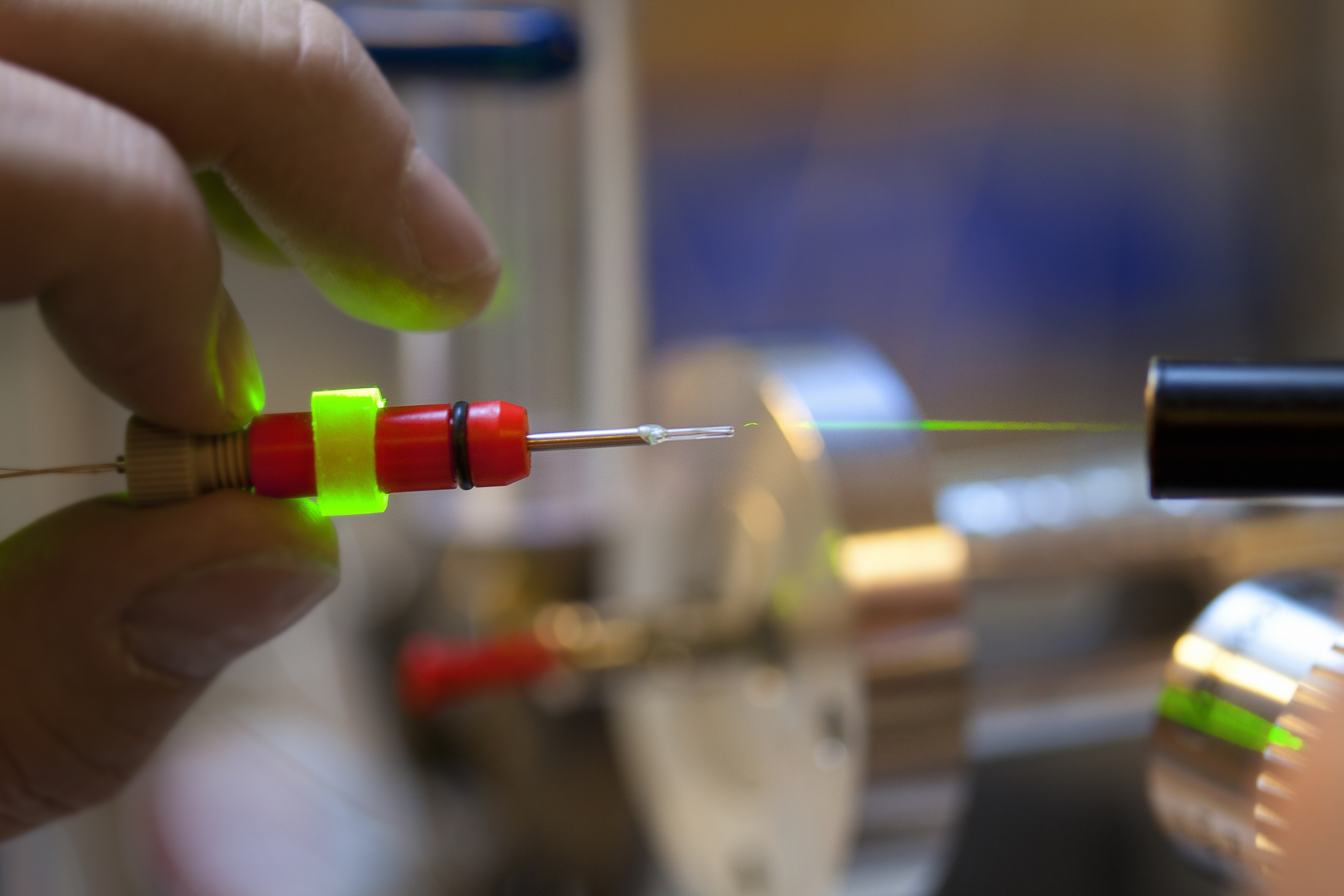
Photo Magnus Bergström
Summary:
Studying non-linear interaction between photons and matter in atoms, molecules and large biological systems.
Developing technologies and science for high-resolution depictions of individual biological molecules, such as virus particles, live single-cell organisms and nanocrystals.