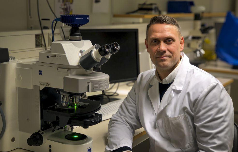
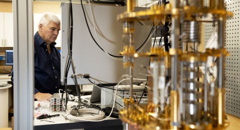
During his first period as a Wallenberg Academy Fellow, Martin Andersson and his research team were the first in the world to analyze tissue using an atom probe. He is now developing a method of determining the exact structure of proteins using the same tool. This may open new doors in drug development.
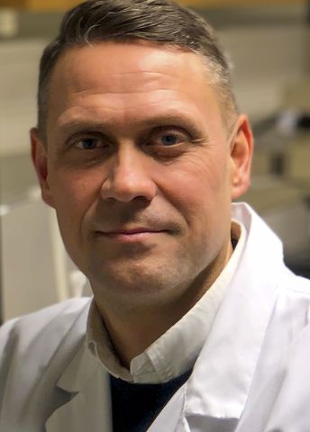
Martin Andersson
Professor of Applied Chemistry
Wallenberg Academy Fellow, prolongation grant 2018
Institution:
Chalmers University of Technology
Research field:
Nano materials and surface chemistry
Mapping protein structure was actually not something that Andersson, who is a professor of chemistry at Chalmers University of Technology in Gothenburg, had planned. For several years he concentrated on creating synthetic bone that enabled implants to knit seamlessly with the skeleton. The need to study the interface between implant and skeleton gave him the idea of trying out a tool not previously used in the fields of biology or chemistry.
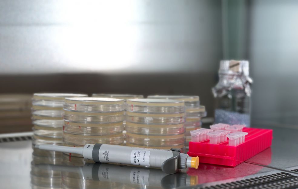
Use of atom probe tomography, as the method is called, entails analyzing a minute, needle-shaped specimen of a material. A strong electric field detaches the atoms, one by one, from the specimen. The time it takes for the atoms to reach the probe’s detector makes it possible to determine their mass and reconstruct their position in the specimen.
“Atom probes have been around for a while, but they’re unusual – the only one in Sweden is here at Chalmers. They were developed for use in metallurgy, and even today their use is virtually confined to the study of metals and other hard materials,” Andersson says.
New use of an established tool
His idea of trying out the atom probe on bone met with skepticism. But he gave it a go nonetheless – and was the first person in the world both to analyze tissue with an atom probe, and to show how the atoms are arranged in the interface between the implant and skeleton. His success whetted his appetite to further explore atom probe potential in biology.
“We analyzed some more bone and included a protein in the specimen. The atom probe enabled us to see the precise structure of the protein, atom by atom. No previous technique for determining structure offers such high resolution.”
Protein structure is central to how proteins work and behave. Many modern drugs target proteins, and it is very useful for drug developers to know more about their structure.
“Imagine if we could develop a method of using the atom probe to examine other proteins as well as those found in bone. It could really help in development of new medicines.”
And thanks to a renewed Wallenberg Academy Fellow grant, that is exactly what Andersson is now doing. Inspired by algae that stabilize proteins using silicon oxide, he and his colleagues have found a method of encapsulating the proteins they want to study in glass. The glass is shaped into ultra-thin needle-shaped specimens, which are then analyzed using the atom probe.
Proteins in the cell membrane are particularly relevant for many drugs. But current techniques for determining structure – X-ray crystallography, MRI and cryo-electron microscopy – often fall short. This is because the structure of membrane proteins is destroyed if they are removed from the membrane.
“The atom probe could be an excellent complement to current techniques. It allows us to let the protein remain in the membrane and encapsulate the whole cell membrane in glass,” Andersson explains.
Tiny needles in a large haystack
The challenge now facing the project is to find the protein embedded in the nano-capsule of glass, to ensure that it is included in the needle-shaped specimen to be analyzed by the atom probe.
“It’s like looking for a needle in a haystack. The nano-capsules are incredibly small – all the atom probe specimens that have been analyzed so far would fit into a single sesame seed.”
So the researchers are now examining various techniques for locating the proteins in the glass fragment, before they begin fabricating specimens. The first step has been to make ultra-thin glass fragments, so they only need to search in two dimensions. Step two will be to make minute indentations in the surface of the glass into which the protein can dock.
“Then we’ll be down to one dimension, and we won’t have to guess where the protein is,” Andersson says.
So far, Andersson’s research team is virtually alone in the world in using atom probe tomography to study the building blocks of biology.
“It’s fun being a pioneer, but also very challenging, since there’s no one to ask if you have a problem. But given the huge potential of atom probe tomography, it feels right to pursue this line of research. And we need funding of the kind offered by the Foundation, with no strings attached.”
“The grant is essential so I can research in this way. It gives me the courage to tackle completely new ideas, and the time to thoroughly investigate new phenomena.”
Andersson’s roots are in chemical engineering, but he has devoted more and more of his energies to biology, and enjoys exploring new avenues. His research into synthetic bone for implants, which gave him the idea of determining protein structure using atom probe tomography, has also given him impetus in other directions.
One example is his work on synthetic cartilage, which could be of benefit in treating osteoarthritis, a widespread condition. And the problem of infections occurring in conjunction with implant surgery has spurred him to design surfaces hostile to bacteria and kind to the body’s cells.
“I’m driven by the fun and creative aspects of research. And I like it when there is a link to real-life problems – where success means helping other people.”
Text Ingela Roos
Translation Maxwell Arding
Photo Mats Hulander