In a part of the brain called the basal ganglia, information is analyzed instantaneously so that our bodily movements and senses work as they should. Gilad Silberberg is studying how the brain organizes this process. Malfunctioning of the system is linked to a number of diseases, including Parkinson’s.
Gilad Silberberg
Professor of Neurophysiology
Wallenberg Academy Fellow, prolongation grant 2017
Institution:
Karolinska Institutet
Research field:
Structural and functional characteristics of the neocortex and the basal ganglia in the brain, and mapping of neuronal activity and interaction in small neural networks
The brain’s network of neurons is a constant hive of activity. Nerve cells of various kinds communicate with each other in advanced interaction – even if we merely take a bite of an apple or wiggle a toe. The brain receives signals from all our senses, from our own body, and from our surroundings. Silberberg elaborates:
“Humans make decisions and transmit information between regions of the brain in milliseconds. And we can react, respond and make decisions simultaneously. It involves incredibly complex logic, which we need to learn more about.”
Huge quantities of information are processed in the striatum, which is the main input structure of the basal ganglia. It functions as a kind of switchboard in the brain, where everything takes place automatically, and normally without us needing to reflect over it.
– Most brain researchers are fundamentally interested in understanding two general functions: how we process sensory inputs and how we generate behavior. These functions are studied in all brain areas, but we focus on a specific region, the basal ganglia. The basal ganglia have been studied mainly in relation to motor functions, and we aim to integrate it with the sensory processing.
Yet this field remains little explored. One reason is inaccessibility. The basal ganglia lie deep at the base of the forebrain under the cerebral cortex.
“Research has long focused on higher brain functions. Less time has been spent on the underlying networks. But in recent years we have begun to close the gap.”
Symphony orchestra
The brain contains over a hundred billion neurons, which form miniature networks. The neurons have special traits and unique functions. They may be likened to the instruments of a symphony orchestra. The cells play the same piece of music but, just as in an orchestra, as different instruments and in separate parts. Silberberg wants to understand how the neurons behave in order to produce concordant music.
“We’re trying to understand the task performed by each instrument in a large orchestra.”
There are now technologies and methods for studying neurons and their activity. There are many questions. Which cells talk to each other, and which do not? Which cells operate at high frequencies and which at low ones? Which cells suppress signals and silence their neighbors, and which ones stimulate other cells and let information pass along neural pathways?
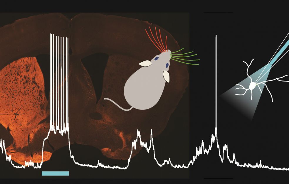
Clues appear all the time. In a series of studies between 2014 and 2020 Silberberg and his colleagues were able to announce new findings about the striatum. It appears that the electric signals from the right and left sides of the body differ, and that neurons of a specific kind in the striatum integrate sensory impressions from the two sides.
The researchers are using a technique known as patch-clamp, which enables them to record synaptic transmission at the contact point between two neurons. The researchers are studying brain activity in mice, and examining the reactions when their whiskers are touched on both sides, and when they are given a visual stimulus.
The results also show that a specific subgroup of neurons in the medial striatum is responsible for integrating sensory impressions from different types, such as touch and light. A new understanding of the basal ganglia is starting to take shape.
“It’s become increasingly clear how motor and sensory functions are intertwined in this part of the brain.”
New findings on Parkinson’s
The basal ganglia are sometimes compromised as a result of diseases and functional impairment. Parkinson’s disease is the best-known example, along with Huntington’s disease, Tourette’s syndrome and ADHD.
For the brain it is crucial to be able to sort impressions coming from the right-hand side from those coming from the left. Silberberg has shown that this function in particular is impaired in Parkinson’s sufferers.
The reason is lower levels of dopamine, a signal substance that plays a key role as a neuromodulator in the basal ganglia. Dopamine causes neurons that are normally experts at distinguishing information from the left- and right-hand sides to lose some of that ability. This may in turn impair the brain’s ability to interpret sensory impressions.
The finding is interesting – not least because it creates entirely new potential for early detection of Parkinson’s disease.
“We now know that Parkinson’s patients can experience a sensory deficit several years before they exhibit impaired motor functions.”
“The Wallenberg Academy Fellow grant represents generous funding, which often reaches researchers at just the right time – when they are at their most productive and creative – while also in need of support. The Fellows network is another asset; network contacts have led to new collaborations.”
Silberberg is working with Johan Lundström – also a Wallenberg Academy Fellow – in a study to ascertain whether an impaired sense of smell can also be used to make an early diagnosis of Parkinson’s disease.
The goal is to pursue this curiosity-driven research, but Silberberg also wants his research findings to be used for medical benefits.
“We have contributed to knowledge about Parkinson’s disease, and I hope that we will become even more engaged in translational research in the future. Many of the findings that lead to better therapies come from research into fundamental functions.”
Text Nils Johan Tjärnlund
Translation Maxwell Arding
Photo Matthijs C. Dorst, Gilad Silberberg