Sten Linnarsson wants to understand how the human brain is formed – from the egg cell to the thousands of cell types found in an adult brain. He is using methods that reveal which genes are active in individual cells to create a detailed map of brain development during the fetal stage.
Sten Linnarsson
Professor of Molecular System Biology
Wallenberg Scholar
Institution:
Karolinska Institutet
Research field:
Molecular neurobiology
“I think it’s incredibly fascinating that the human brain, which is so complex, is formed from a single cell, and builds itself,” says Linnarsson, Professor of System Biology at Karolinska Institutet.
Like other bodily organs, the brain’s development begins with a fertilized egg cell. That cell divides into more cells, which change form and characteristics as they multiply before eventually forming the thousands of different cell types that make up an adult brain. Linnarsson, who is a Wallenberg Scholar, is examining how the process works.
“We want to identify the order in which this huge multitude of cells is formed, and how it is done. We will be studying development of the human brain during the fetal stage in much greater detail than anyone has done before.”
“The grant enables me to undertake ambitious, risky projects. It gives me the freedom to pursue research wherever it leads, and follow new, promising leads as they arise. In practical terms, it enables us to establish a platform for studying embryonic development of the human brain.”
Examining individual cells
To achieve this, the research team is using a method of identifying the genes that are activated in individual cells.
All our cells contain the same DNA, the same genes. When different types of cell are formed, certain genes are activated in some cells, other genes in others. This gives the cells their specific functions. The researchers measure gene activity in individual cells. This enables them to analyze cells in greater detail, and obtain a picture of the different cell types in a given organ.
Linnarsson is a pioneer in the field of single-cell analysis. In 2011 he showed that it is possible to measure gene activity in hundreds of individual cells simultaneously in a single sample. Progress since then has been swift.
“Now we can analyze up to a million cells in a single experiment – the cost per cell has fallen dramatically. This enables us to study much more complex tissues than was originally the case.”
Active genes form RNA, a working copy of DNA. Linnarsson has developed a method of counting RNA molecules in a cell. This makes single-cell analyses more accurate.
Heart of the method
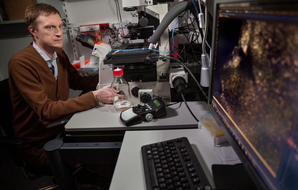
On a laboratory bench at the Biomedicum research center lies a black box, about the size of a shoe box. It is not much to look at, but is the machine that makes the study possible.
“It encapsulates cells from a tissue sample one by one in droplets of fluid, where the reactions take place.”
Each droplet contains one cell, certain reagents and a unique fragment of DNA. The cell’s RNA is transcribed to DNA in the droplet and combines with the unique DNA fragment.
DNA from all droplets can then be sequenced simultaneously. This is because the DNA fragment enables the scientists to identify which RNA sequences come from the same cell, i.e. which genes are active in the cell.
During the project the researchers will analyze millions of embryonic cells from week five to week twelve after fertilization – the period when the fundamental brain cell types form. The result will be a development tree showing how specialized nerve cells are created, and which genes are activated in them.
The researchers can also monitor changes in gene activity in the cell, using a method developed by Linnarsson and fellow researchers in the U.S. It exploits the fact that when a gene is activated, an unedited RNA molecule forms first, before being modified to form mature RNA. The quantity of those molecules is compared, enabling the researchers to identify the stage of the modification process that the cell has reached.
“We can see the genes that are active now, as well as the state of the cell a few hours previously. Many paths taken by cells during embryonic development are chosen over this time scale.”
The aim is to create a mathematical model capable of predicting which genes drive the cell in a given direction. This can then be used to predict the consequences of abnormal changes, such as destruction of a specific gene.
Culturing brain tissue
Linnarsson draws the black drape at the entrance to the microscopy room to one side. Cells show up as dots on a computer screen.
“It shows a section of tissue from an embryonic brain. We have stained a subset of the cells so we can distinguish them.”
The scientists can keep fragments of embryonic brain tissue alive in the lab for some weeks. They are cultured under a microscope connected up to a camera. The researchers stain specific cell types so they can see how the cells mature and move, and test the predictions made by the mathematical model.
“The next step is to knock out a gene to see whether a cell that would have become an astrocyte, for instance, becomes something else instead. But there’s still some way to go before we can do this.”
They are also staining individual RNA molecules in frozen tissue sections to see where the active genes lie. This may be of central importance to how the proteins formed by the genes impact the cells around them.
Values freedom
Linnarsson explains that he became a researcher because he has always been curious to know how things work. His interest in the brain was awakened when he studied molecular biology, and he received a PhD in neurobiology at Karolinska Institutet.
He then left university to start a company based on a new method of DNA sequencing. But he always planned to return to research.
“Here I have the freedom to pursue the scientific path I find interesting without anyone deciding what I should do. It’s a challenging job, but one I really enjoy.”
Text Sara Nilsson
Translation Maxwell Arding
Photo Magnus Bergström