Hans Hertz’s choice of career was by no means a foregone conclusion. He tried both law and business before opting for physics. Now he is the driving force behind one of the greatest innovations in the X-ray field since the 1930s. But the technique is yet to be used on humans.
Hans Hertz
Professor of Biomedical Physics
Wallenberg Scholar
Institution:
KTH Royal Institute of Technology
Research field:
X-ray physics
Hertz says that the goal has always been to see smaller things than we can today:
“Ideally, I’d like to be able to see inside a cell in the brain to see what its functions are – which proteins the cell expresses.”
This is, to say the least, an ambitious target for a physicist. That is why he has put together an interdisciplinary research environment in which physicists are working shoulder to shoulder clinicians, biologists and chemists. They are collaborating so closely that for a time they were thinking of transferring all technology to Karolinska Institutet. But it was instead decided to develop the environment at the AlbaNova University Centre.
“We now have clinicians working with us who know much more about medical issues and cell biology than we do. With everyone doing what they are best at, we hope the result of one plus one will be a little more than two.”
From mice to men
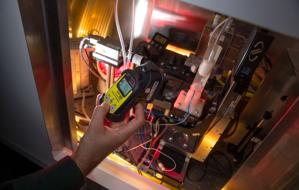
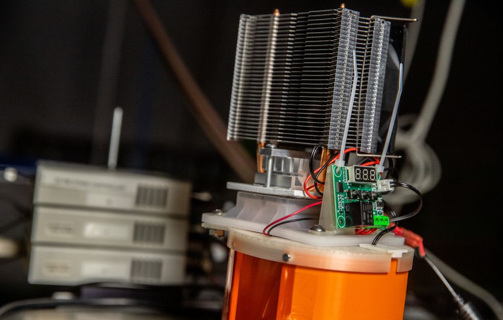
Achieving the goal of imaging the infinitesimal has required a quantum leap in technology – the development of a new X-ray method that produces sharper images, better contrasts, and requires shorter exposure times. The innovation is called liquid metal jet technology. Simply put, it involves replacing the positive anode in an X-ray tube with a jet of liquid metal. This makes it possible to dramatically increase the power output without any risk of overheating.
“Now we can see cells at a size of 0.01 millimeters in a sample that’s about one centimeter across. The next step is to see individual cells in a living mouse. What we learn there should enable us to take the next step – human beings.”
Simulations have already been made, but the next step requires collaboration with clinical researchers, whose role will include helping to interpret the images.
“We’re pretty good at physics and technology. Now we’re learning more about the constraints imposed by biology, and over the next year we’ll be bringing in more chemists.”
Mummies in the lab
Hertz has received generous funding from Knut and Alice Wallenberg Foundation over the past few years. Having been chosen as a Wallenberg Scholar, his grant has now been extended.
“We’ve spent the past five years broadening our palette. Today we have a nanochemistry lab, a good cell biology lab, where we can also carry out virus experiments, and our own facility for animal experiments. We wouldn’t have been able to build up this structure without the Scholar grant.”
A somewhat unexpected application of the X-ray method was reported just over a year ago. During a spell overseas Hertz met Salima Ikram, a professor of Egyptology at the American University in Cairo.
“She showed me her X-ray pictures of mummies, and I said we could improve on them. It then turned out that she kept her catalog of mummies at the Museum of Mediterranean and Near Eastern Antiquities in Stockholm.”
A mummified hand was packed and carefully transported to the lab for examination. The resulting images showed soft tissue, particularly fat cells, at a higher resolution than ever before.
“We discovered there was a field of science called paleopathology, which is the study of disease through the ages. X-ray imaging is a central tool in that field, but to date they have only been able to see bones and teeth,” Hertz explains.
Publication of the X-ray images of the 2,400-year-old mummy was front-page news in the X-ray journal Radiology.
“Now we’re looking for a killer application for the field, and the PhD student who performed the study, Jenny Romell, will be attending conferences to make contacts.”
Commercialization – a key tool
Liquid metal jet technology underpinned a spin-off – Excillum – a company that is now refining the technology. The road to commercialization provided a unique opportunity to gain an insight into a new field. Hertz elaborates:
“And if I really want to see change, I have to make a product. Only then will the technology be of any benefit. And as long as I am not involved in the company’s operations, I can keep the two roles apart.”
The first step in creating the liquid metal jet technology was an X-ray source based on laser plasma. The idea was also to spin off that discovery as a separate company. Hertz explains:
“But at the time we were too inexperienced to successfully commercialize the technology ourselves. Others have now taken up the reins, and made it possible to make much smaller microchips.”
Time-consuming cross-border collaboration
In recent years much time has been spent creating a fertile culture. Hertz thinks that learning to collaborate between disciplines is far from simple:
“It takes a long time to get it to work, and to develop a research climate in which we understand each other and can benefit from our respective strengths. It would have been really hard to achieve without the Wallenberg funding.”
Thanks to the Wallenberg Scholar grant it will now be possible to obtain X-ray images from a live mouse with details at a scale of ten micrometers – and a complete calculation of how this can be done in humans.
“Then we can begin the next journey – assuming we obtain funding, and judge that it can be done. But I am hopeful.”
Text: Magnus Trogen Pahlén
Translation: Maxwell Arding
Photo: Magnus Bergström