A new generation of electron microscopes has enabled researchers to examine the structure of proteins found in cell membranes in a more detailed and more efficient way. Better knowledge about these proteins may lead to better drugs.
Project Grant 2014
Single-particle cryo-EM studies of membrane protein biogenesis and structure
Principal investigator:
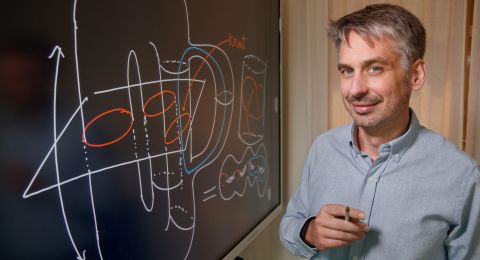
Gunnar von Heijne, Professor of Theoretical Chemistry
Institution:
Stockholm University
Grant in SEK:
SEK 31 million over five years
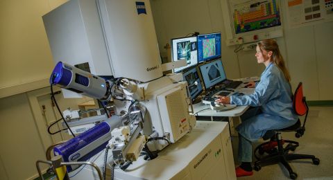
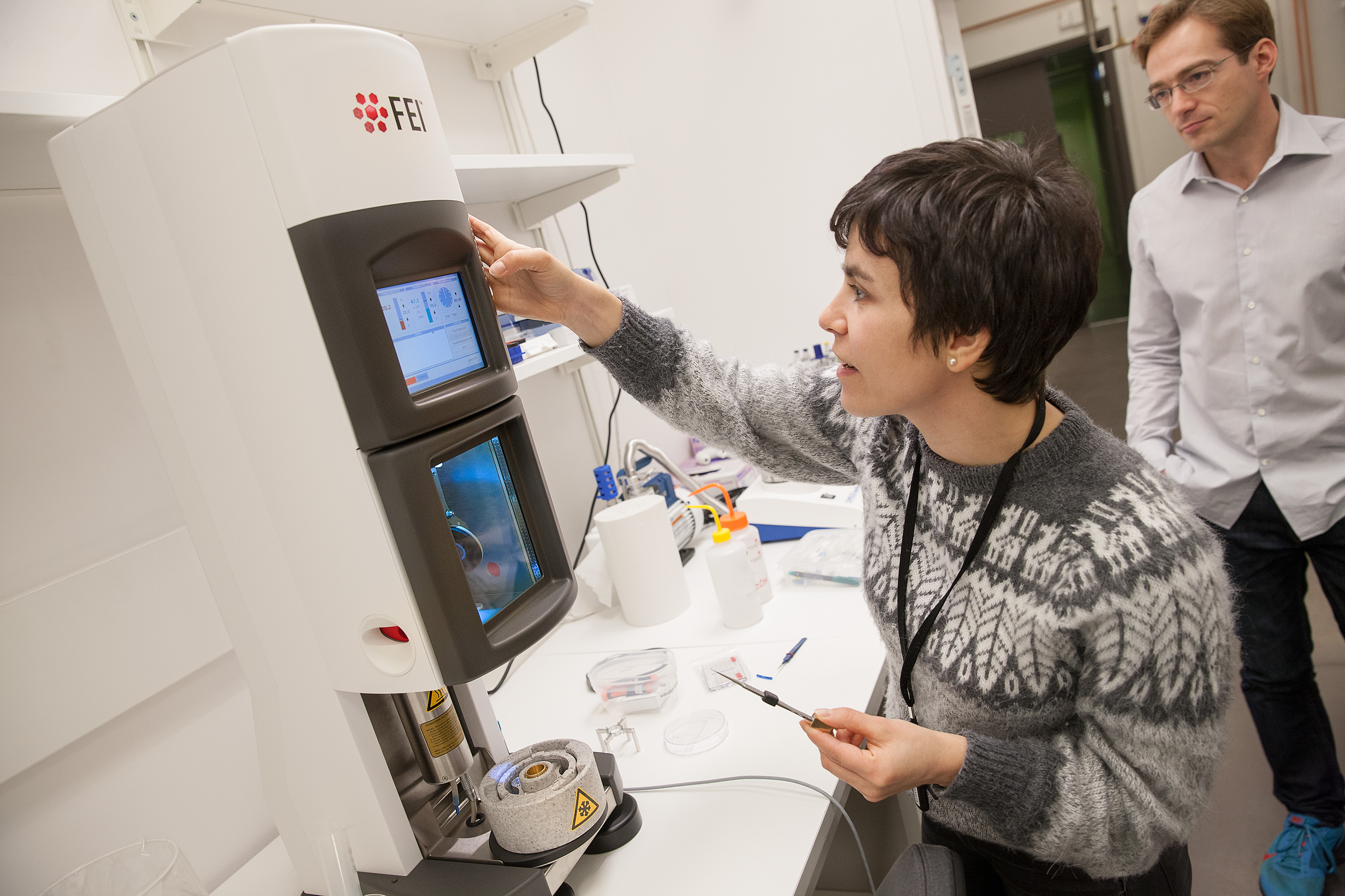
Gunnar von Heijne, Professor of Theoretical Chemistry, was the driving force behind the purchase of an advanced “cryo-electron” microscope for studying single particles. He explains how luck was on his side when the microscope was delivered to SciLifeLab in Solna, north of Stockholm:
“We were pretty nervous – it weighed two metric tons, and we only just got it into the elevator.”
The microscope is fitted with a new kind of electron detector that makes it easier and quicker to determine the structure of larger proteins and protein complexes, including the type of proteins found in the protective membrane of cells.
“There is an enormous amount of interest in using the microscope. A great deal of preparation has been necessary, and we hope to bring the facility into operation in summer 2016.”
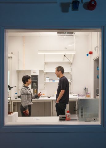
The microscope will only work perfectly if the ventilation and cooling are up to standard. Four rooms on the first floor at SciLifeLab have been specially equipped for the purpose. There is also a second, smaller electron microscope and various equipment needed to prepare and freeze samples.
Membrane protein structure
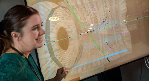
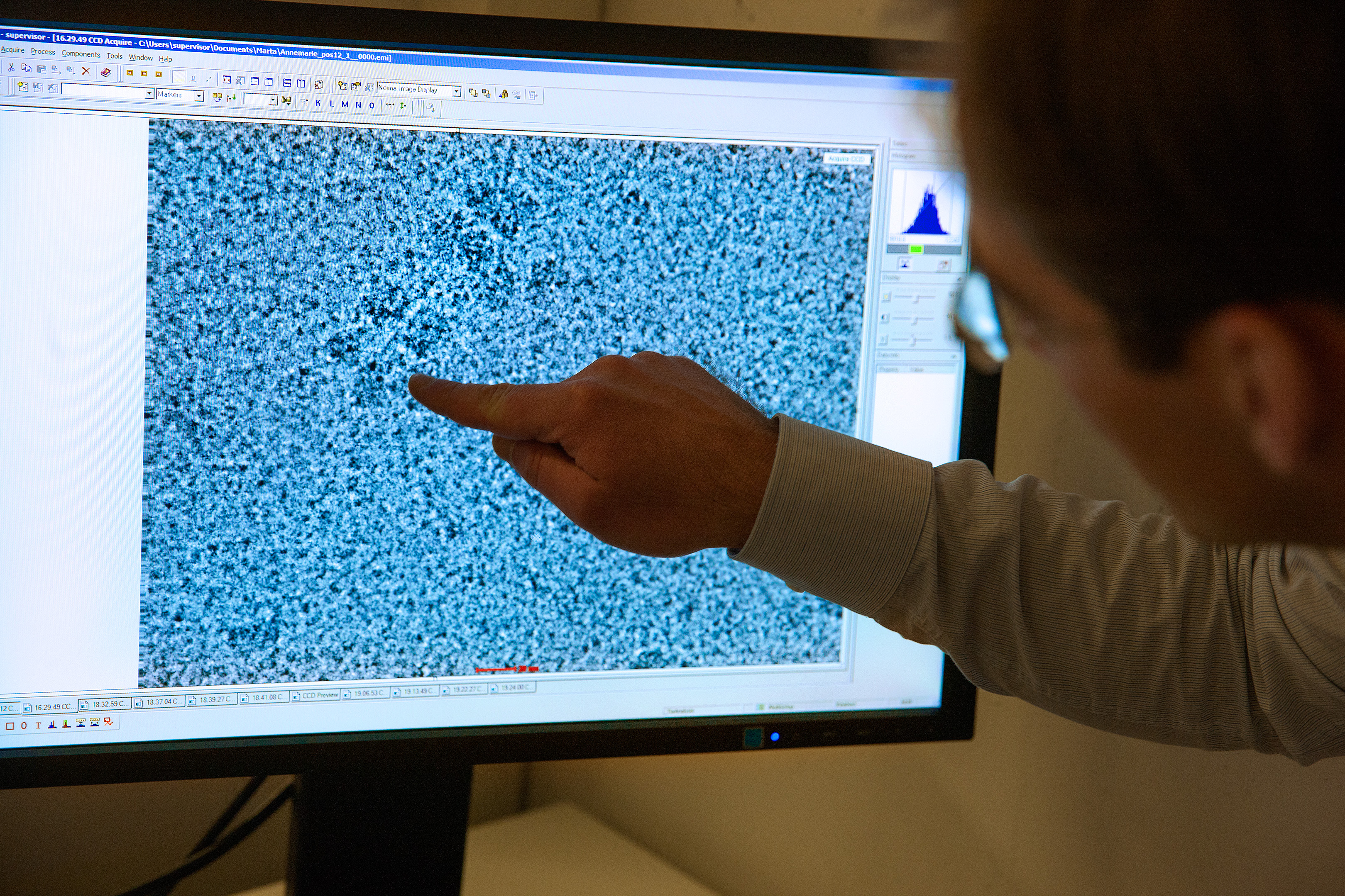
First, the protein molecules imaged in the cryo-electron microscope are frozen into a thin film of ice with the help of liquid nitrogen.
“Using the images of tens of thousands of individual molecules produced by the microscope, we can then work out the three-dimensional structure of the protein on the computer. Huge quantities of data are involved.”
Professor von Heijne’s interest in cryo-electron microscopy is rooted in many years’ successful research into membrane proteins. He heads the Center for Biomembrane Research at Stockholm University, which is a center of excellence in the field.
One-third of a cell’s proteins are found in the protective lipid membrane surrounding it. Membrane proteins have many vital functions, and knowledge of their structure and characteristics is a key factor in the development of new drugs. In fact, just over half of all current medicines target membrane proteins.
“Traditionally, we use X-ray crystallography to determine the structure of proteins. We try to get the protein to form crystals, which we illuminate with a powerful X-ray beam. But membrane proteins are hard to crystallize.”
With cryo-electron microscopy it is not necessary to make crystals to determine the structure. The only limitation in using this technology is that the proteins must be large enough, as Professor von Heijne explains:
“It doesn’t work with really small proteins – there we have to use crystallography, but the limit is being extended ever downwards.”
Timing and collaboration
It was pure luck that cryo-electron microscopy caught Professor von Heijne’s attention in late 2013.
“I usually spend a month at Rockefeller University in New York in the fall. I have a colleague there who is a structural biologist specializing in membrane proteins, just like me. That year there had just been a breakthrough for electronic imaging detectors, and membrane proteins were among the subjects being studied, since they are so hard to crystallize. My colleague saw the potential for using this technology in our research.”
The microscope has been funded jointly by the Knut and Alice Wallenberg Foundation and the Erling-Persson Family Foundation. Half the time it will be available as a national resource. The rest of the time it will be used by researchers in the project to determine the structure of membrane proteins, among other things.
The Knut and Alice Wallenberg Foundation has also contributed to a similar facility at Umeå University.
“At Umeå they will mainly be using the microscope for cryo-tomography, which involves studying cell sections instead of single-particle cryo, which is what we do. They have the same arrangement, that the facility will be a national resource half the time. So it was natural to coordinate this under the SciLifeLab umbrella.”
Ribosomes under the microscope
The research is taking place in close collaboration with other leading international research teams at Ludwig-Maximilians-Universität, LMB-MRC in Cambridge, Rockefeller University and Stanford University, which all use this technology.
One line of research where the new microscope may be of great benefit concerns the way proteins fold when they are synthesized from ribosomes – enzymes that make proteins.
“When the protein has reached a certain size, part of it may begin to fold, even though it remains attached to the ribosomes. We have studied this in a number of samples using single-particle cryo at Ludwig-Maximilians-Universität in Munich. We were the first to do so.”
The researchers will also be using the microscope to study ribosomes and biogenesis of membrane proteins in the mitochondria – the powerhouses of the cells.
“This technology allows us to use much more imagination in the design and content of the samples. I think the most interesting aspect of this is the scope offered to build and study larger complex molecules.”
Text Susanne Rosén
Translation Maxwell Arding
Photo Magnus Bergström